The skeletal system
Apréndelo jugando
Responde estas preguntas para ganar energía, luego pesca y explora. Sin cuenta.
Para educadores: diapositivas de la lección, apuntes de repaso listos para usar sobre The skeletal system (Physical Education, Anatomy and Physiology) — úsalos en tu lección, o presenta el tema como una actividad interactiva de clase que tus aprendices juegan como un juego en vivo.
Apuntes de la lección
Functions of the skeleton
- The skeleton supports the body, provides muscle attachments and acts as a system of levers around joints. Cranium helps protect the brain and the thoracic cage helps protect heart and lungs. The muscles supply active force; bones do not contract to produce movement.
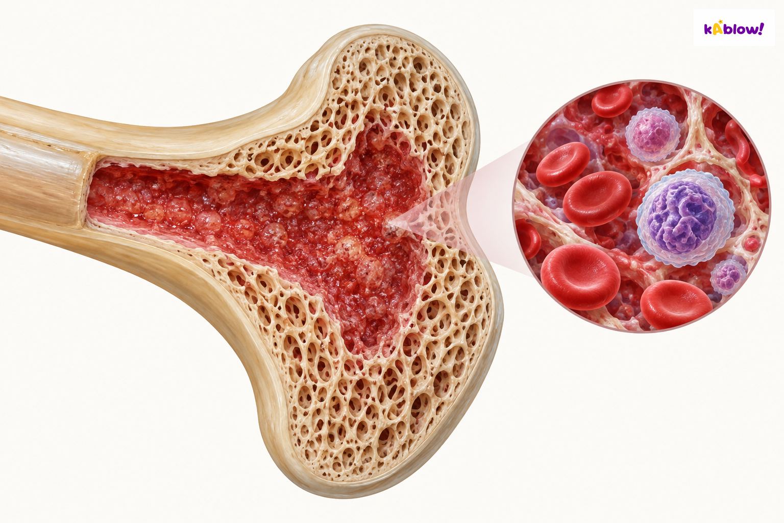
- Red marrow forms blood cells; red cells carry oxygen, white cells support immune defence and platelets contribute to clotting. The illustration shows a marrow concept, not the precise adult distribution: red and yellow marrow locations change with age. Calcium-containing mineral supports bone strength and forms a regulated mineral reservoir.
Red marrow and blood-cell formation
Main bones
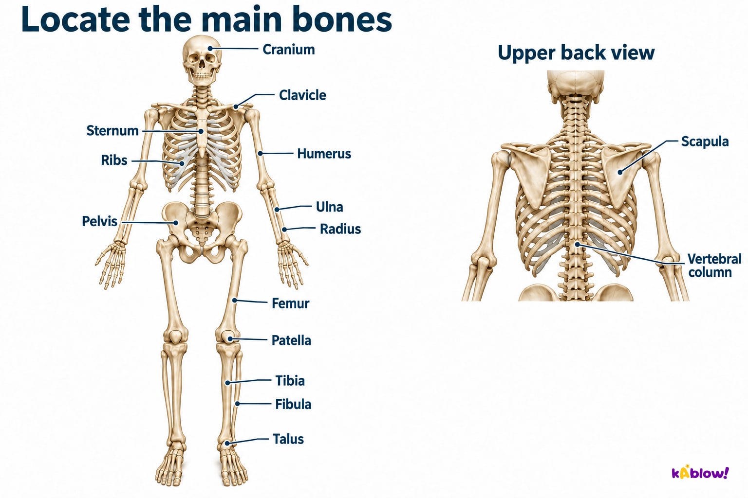
- Use anatomical position, with palms forwards, to identify radius on the thumb side and ulna on the little-finger side. Humerus is in the upper arm; clavicle crosses the upper front chest, scapula lies at the back and sternum is midline among the ribs. The chart’s posterior inset makes scapula and vertebral-column locations clearer.
- Pelvis connects trunk and lower limbs; femur is thigh, patella lies in front of the knee, tibia is the large medial shin bone and fibula the slender lateral bone. Talus is a tarsal beneath tibia at the ankle. The figure labels selected main bones rather than every bone of the skeleton.
Locate the main bones
Types of bone
- Bone type describes shape, not simply size or function. Femur and humerus are long bones, and so are small finger phalanges. Carpals and tarsals are generally short bones; ribs, sternum and most cranial-vault bones are flat. The cranium is made of several bones and is not one uniformly flat bone.
- Vertebrae have complex irregular shapes; irregular is an anatomical category, not damage. Patella is a sesamoid bone embedded in a tendon. Categories can support different functions, but a long bone can also contain marrow and a flat bone can provide muscle attachments.
Classify by shape
Joints
- A joint is where bones meet. Hinge examples chiefly allow flexion/extension in one plane; ball-and-socket hip/shoulder permit movements about several axes within limits. The atlanto-axial pivot contributes to head rotation. Knee is a modified hinge and the elbow region includes radius-ulna pivot articulations as well as its main hinge action.
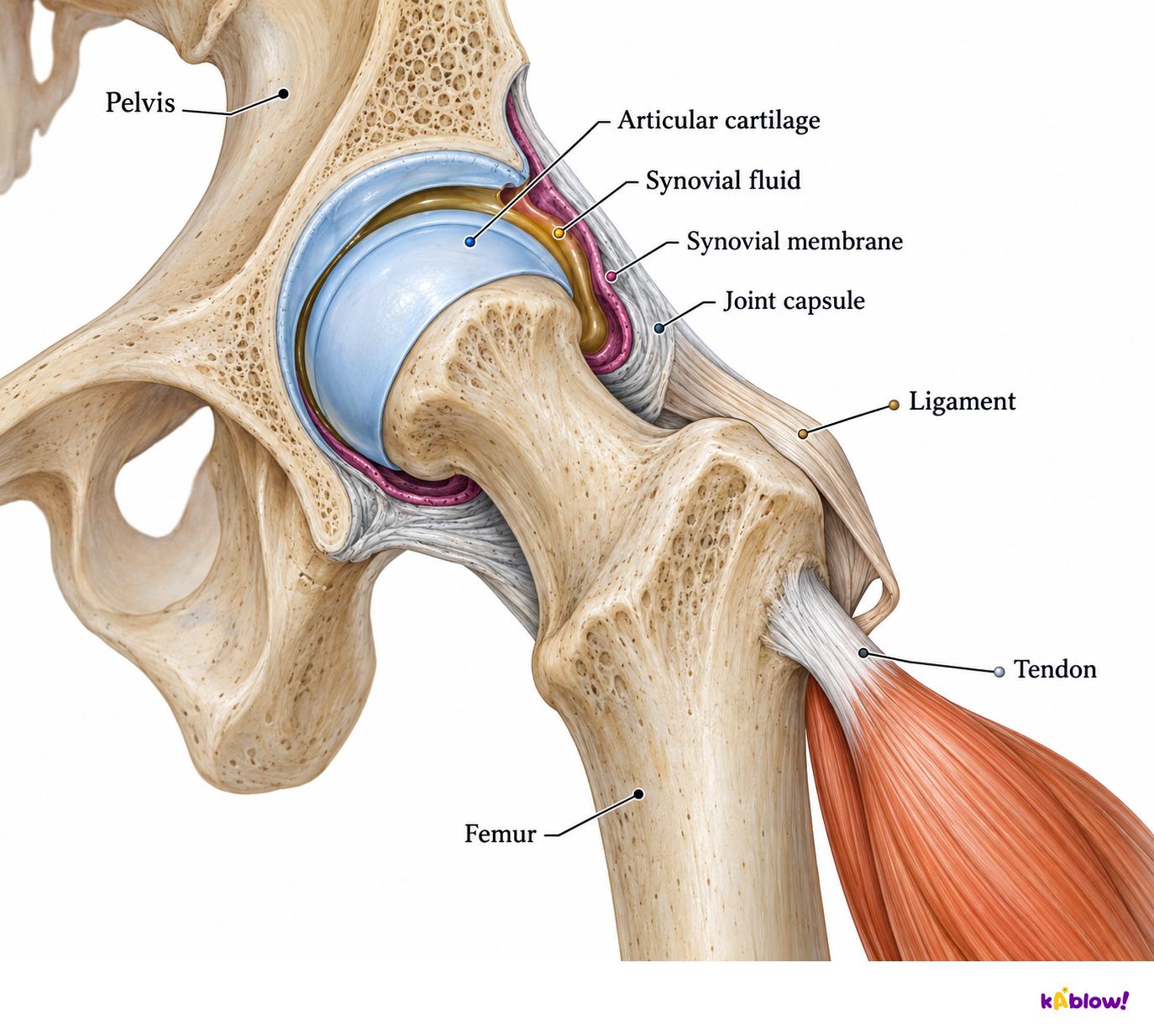
- The hip cutaway shows articular cartilage on bone surfaces, synovial fluid in the cavity, a fluid-producing synovial membrane and outer capsule. Ligaments connect bone to bone; tendons transmit muscle force to bone. Cartilage and fluid reduce friction and help distribute loads. Bursae are additional fluid-filled sacs reducing friction between nearby tissues, not the main joint cavity, and are not drawn in this image.
A synovial hip joint
Movements
- Flexion decreases the joint angle and extension increases it in typical knee/elbow examples. Shoulder abduction moves the upper arm away from the midline; adduction returns it towards the trunk. Specify the joint and direction rather than describing only a whole skill, since several joints can move differently in one action.
- Rotation turns a bone about its longitudinal axis. Circumduction combines flexion, abduction, extension and adduction so the limb sweeps a cone; it is not the same as axial rotation. Ankle dorsiflexion draws the front of the foot towards the shin; plantar flexion points it away. Bone shape and supporting tissues limit each movement range.
Describe movement at a named joint
Posture
- The vertebral column supports the trunk and helps protect the spinal cord. Muscles, joints, discs and ligaments contribute to movement and support. People have different spinal curves and comfortable positions, so there is no single ideal posture that everyone must hold.
- A visible posture alone does not diagnose a cause of pain. Task demands, comfort and many individual factors matter; varied comfortable positions and suitable activity can support function. Persistent symptoms call for individual assessment rather than assuming a person’s body shape or one position is inherently faulty.
Support does not imply one perfect posture
Bones at named joints
- Shoulder: humerus meets scapula; hip: femur meets pelvis. Elbow complex: humerus, radius and ulna, with hinge-like flexion/extension and nearby pivot movement for forearm rotation. Knee: femur and tibia with patella at the front; the fibula is nearby but is not the principal femur-contacting bone.
- At the main ankle joint, tibia and fibula form a mortise around the talus. Dorsiflexion and plantar flexion occur here. Inversion/eversion involve other foot joints and must not simply be labelled the same ankle-hinge action. These are selected joint relationships rather than a complete anatomical atlas.
Bones meeting at selected joints
Locate hand and foot groups
- Carpals form the wrist, metacarpals lie in the palm and phalanges form fingers. Tarsals include talus in the ankle/hindfoot region, metatarsals extend through the forefoot and phalanges form toes. Similar naming does not mean wrist bones and ankle bones are interchangeable.
- Location grouping and shape classification are different. Finger phalanges are small long bones; carpals are generally short bones. The patella at the knee is a sesamoid bone in a tendon. Identify the structure before assigning its shape category.
Hand and foot groups
Connect structure to a sporting action
- During a runner’s knee bend, the knee flexes as the angle between thigh and lower leg decreases. Femur and tibia provide the main articulating bones and patella lies in front. Tendons transmit muscle force and ligaments help control the available range; cartilage and synovial fluid support low-friction loading.
- An ankle drawing the foot towards the shin is dorsiflexion at the tibia/fibula/talus joint. The direction identifies the movement, but the active contraction also depends on load: muscles can shorten, lengthen under tension or hold length. Do not assume every straightening or lowering action is produced by a shortening extensor.
Analyse structure and action together
Diapositivas
Sign up free to view the lesson slides
Step through every slide for this topic — plus flashcards and revision notes — with a free account.
Preguntas de práctica
Vista previa gratis — 8 de 40 preguntas. Regístrate para verlas todas.
1.Which named part of the skeleton encloses and helps protect the brain?
Easy- Acranium
- Bfemur
- Chumerus
- Dsternum
2.Which bones protect the heart and lungs?
Easy- Afemur
- Bradius
- Ctibia
- Dribs
3.Which bone is the shin bone?
Easy- Afemur
- Bulna
- Ctibia
- Dfibula
4.Which bone is the upper arm bone?
Easy- Aclavicle
- Bhumerus
- Cradius
- Dulna
5.Which bone is the kneecap?
Easy- Apatella
- Bfemur
- Ctibia
- Dtarsal
6.Which bone is the collar bone?
Easy- Ascapula
- Bsternum
- Chumerus
- Dclavicle
7.Which bone is the shoulder blade?
Easy- Ahumerus
- Bradius
- Cscapula
- Dclavicle
8.Where are red blood cells produced?
Easy- Acartilage
- Bred bone marrow
- Cmuscle
- Dskin
Unlock all 40 questions & more
Crea una cuenta gratis para ver todas las preguntas, las diapositivas, las tarjetas y los apuntes de repaso de este tema.
Exámenes anteriores
La práctica con exámenes anteriores de este tema llegará pronto.
Próximamente