
The cardiovascular and respiratory systems
邊玩邊學
回答這些題目賺取能量,接著就能釣魚、探索。不需要帳號。
給教育者: 為 The cardiovascular and respiratory systems(Physical Education、Anatomy and Physiology)準備好可直接使用的課程投影片, 複習筆記——用於你的課程,或把這個主題當成互動班級活動,讓學習者以即時遊戲的方式進行。
課程筆記
The heart
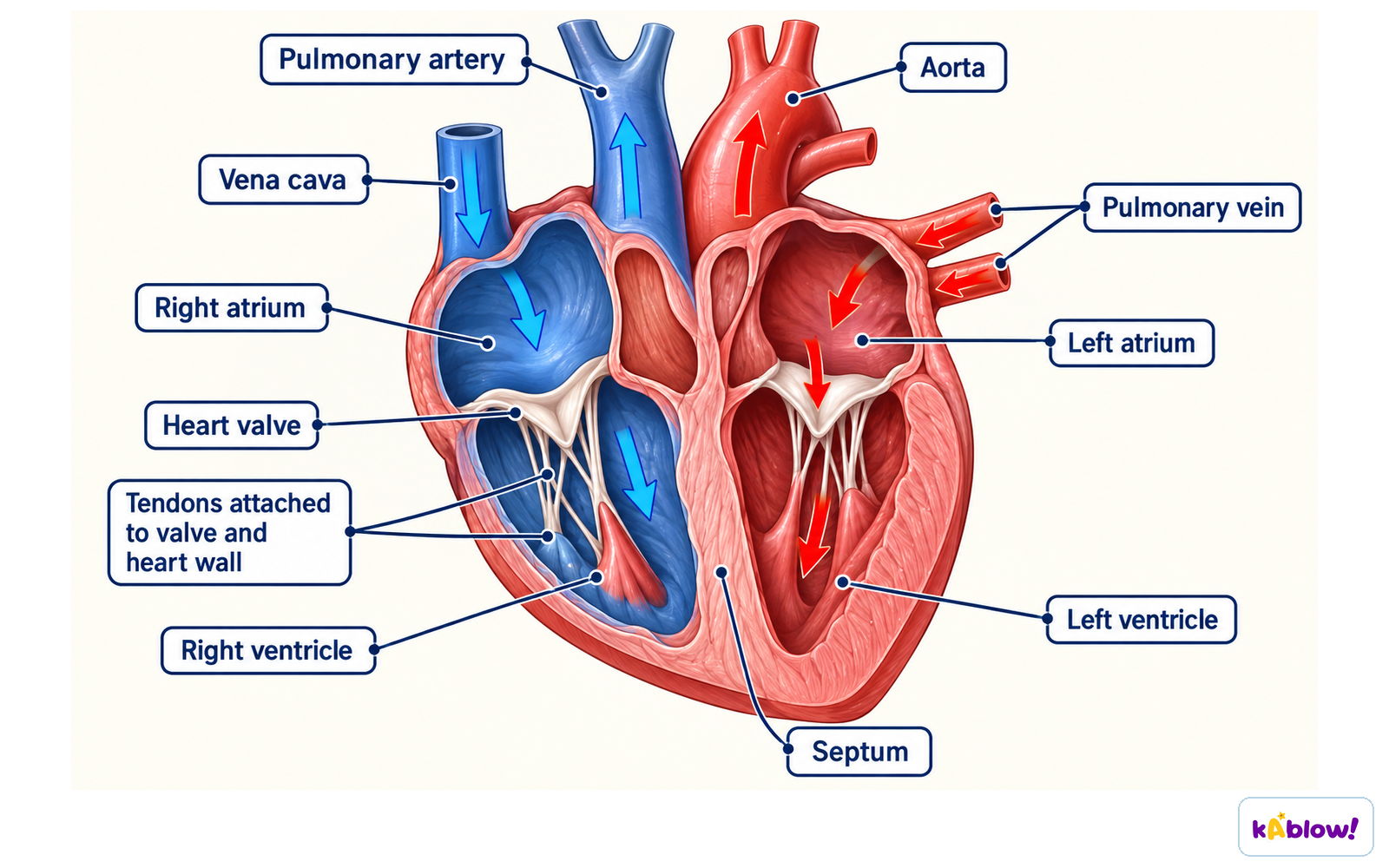
- Four chambers organise a double circulation: venae cavae → right atrium → right ventricle → pulmonary arteries → lungs → pulmonary veins → left atrium → left ventricle → aorta → body. Atria receive and ventricles eject, with the septum separating sides and pressure-operated valves preventing backflow. Anatomical right is on the viewer’s left in the front-view image; blue indicates oxygen-poor blood symbolically.
- Heart rate is beats/min and stroke volume the volume ejected by a ventricle per beat. Their product is that ventricle’s cardiac output; do not add the two sides. Chamber systole concerns contraction and ejection, while diastole concerns relaxation and filling, with coordinated pressure changes opening and closing valves.
Heart chambers, major vessels and one-way flow
Blood vessels
- Arteries carry blood away from the heart, veins towards it. They are defined by direction rather than oxygen content: pulmonary arteries carry oxygen-poor blood to lungs and pulmonary veins return oxygen-rich blood to the left atrium. In systemic circulation the aorta carries oxygen-rich blood towards tissues.
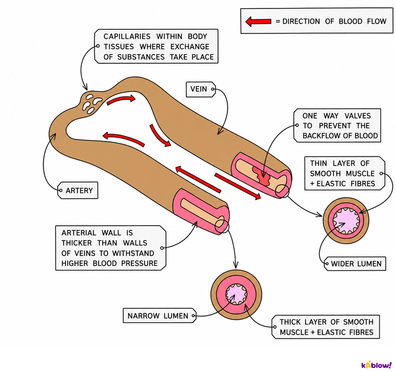
- Comparable arteries generally have thicker muscular and elastic walls for higher-pressure flow, while veins have thinner walls and often wider lumens; many limb veins contain valves limiting backflow. Capillary endothelial walls are thin for exchange. The artery-vein drawing is schematic, and not every vein contains valves.
Artery and vein structure supports different pressures
Blood
- Red blood cells contain haemoglobin, binding oxygen in the lungs and releasing it towards tissues. White blood cells contribute to immune defence through several mechanisms, and platelets are fragments contributing to clot formation alongside plasma proteins.
- Plasma is the liquid transporting cells and dissolved substances such as nutrients, hormones and much carbon dioxide in chemical form. The components have linked functions; red cells are not the only form in which every transported substance travels.
Blood components have linked roles
Vascular shunting
- Vascular shunting is regulated redistribution of blood flow according to demand. Vasodilation widens vessels supplying active tissues, while selected vasoconstriction can reduce the share reaching less active regions. Changes in cardiac output and local regulation also matter.
- Do not describe it as all blood leaving every other organ or the heart making new blood instantly. Skin flow also changes with heat-loss needs, so workload and environmental conditions influence the distribution.
Vessel changes redistribute flow
The respiratory system
- The airway route is nose or mouth, trachea, bronchi, smaller bronchioles and alveoli. Conducting airways move air towards exchange spaces; blood vessels belong to the circulation and are not part of the air route. The flow chart is an order diagram, not a body shape.
- Ventilation moves air, gas exchange transfers oxygen and carbon dioxide across the alveolar-capillary barrier, and circulation transports them. Separating these processes helps explain how the respiratory and cardiovascular systems work together to supply working muscles.
Air route towards alveoli
Breathing
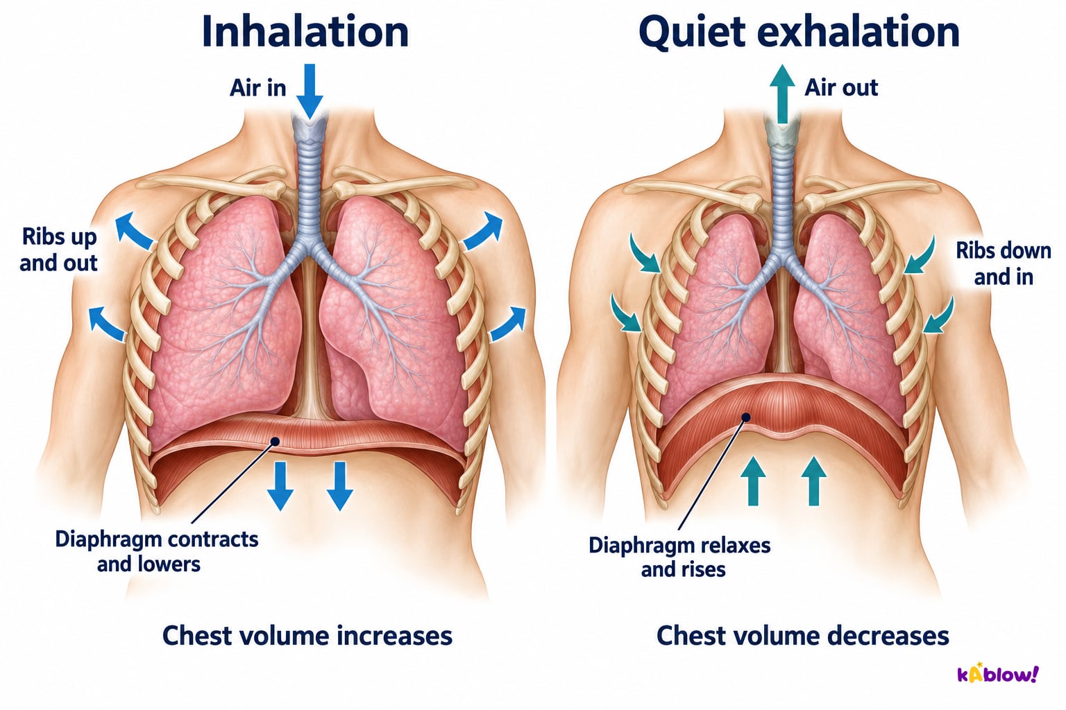
- During normal inspiration the diaphragm contracts, lowers and flattens while external intercostals raise the ribs up and out. Chest volume increases and pressure falls below atmospheric pressure, producing airflow in. The new paired figure shows the lower flatter diaphragm on the left and the higher dome on the right, with leaders ending on muscle.
- During quiet expiration the diaphragm and external intercostals relax and elastic recoil decreases chest volume; pressure rises relative to atmosphere so air leaves. Forced expiration recruits additional muscles, including abdominal and internal intercostal action. Quiet and forced breathing must not be treated as identical.
Inhalation and quiet exhalation
Lung volumes
- Tidal volume is air in one breath under the stated conditions. Inspiratory reserve is extra air inhaled beyond an ordinary inspiration, expiratory reserve extra air exhaled beyond an ordinary expiration, and residual volume remains after maximum expiration. Vital capacity combines tidal and reserve volumes and excludes residual volume.
- Minute ventilation is breathing rate multiplied by tidal volume, giving air volume per minute. It is not vital capacity or oxygen uptake; ordinary spirometry does not directly measure residual volume. Definitions matter more than treating one fixed lung volume as a normal value for every learner.
Distinguish lung-volume terms
Alveolar gas exchange
- Many alveoli create a large exchange area; thin alveolar epithelium and capillary endothelium create a short diffusion distance. Oxygen moves from alveolar air towards blood and carbon dioxide in the opposite direction, down their respective gradients. Haemoglobin binding supports oxygen transport after diffusion.
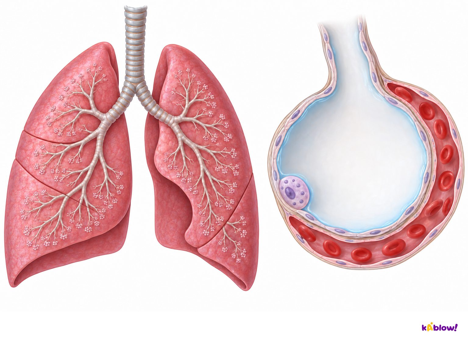
- The existing raster shows lungs and branching airways on the left and an enlarged air space with surrounding red-cell-containing capillary on the right. Ventilation and blood flow maintain gradients; gases cross the thin barrier rather than air travelling along blood vessels. The illustration is schematic and magnified, not to scale.
Branching lungs and the thin alveolar-capillary barrier
Blood output and air ventilation calculations
- Fictional heart rate 60 beats/min and stroke volume 70 mL/beat give output 4,200 mL/min, or 4.2 L/min. Multiply rather than add, and convert millilitres to litres by dividing by 1,000. Cardiac output concerns one ventricle rather than the sum of both.
- Fictional breathing rate 20 breaths/min and tidal volume 0.8 L/breath give minute ventilation 16 L/min. This is air movement, not oxygen uptake or blood output. The inputs are original teaching examples rather than personal reference values or training targets.
Fictional blood and air calculations
投影片
練習題
免費預覽——39 題中的 8 題。註冊即可查看全部。
1.How many chambers does the heart have?
Easy- Afour
- Btwo
- Cthree
- Dsix
2.Which vessels carry blood away from the heart?
Easy- Aveins
- Bcapillaries
- Cnone
- Darteries
3.Which vessels carry blood to the heart?
Easy- Acapillaries
- Bnone
- Cveins
- Darteries
4.Where does gas exchange occur in the body tissues?
Easy- Athe heart
- Bcapillaries
- Carteries
- Dveins
5.What do valves in the heart do?
Easy- Aprevent backflow
- Bpump blood
- Cmake blood
- Dstore oxygen
6.Which gas is carried by haemoglobin in red blood cells to working tissues?
Easy- Aantibodies
- Bclotting factors
- Chormones only
- Doxygen
7.What do white blood cells do?
Easy- Aclot blood
- Bcarry carbon dioxide only
- Cfight infection
- Dcarry oxygen
8.What do platelets do?
Easy- Adigest food
- Bhelp blood clot
- Ccarry oxygen
- Dfight infection
歷屆試題
這個主題的歷屆試題練習即將推出。
即將推出