Animal tissues, organs and organ systems
Apréndelo jugando
Responde estas preguntas para ganar energía, luego pesca y explora. Sin cuenta.
Apuntes de la lección
Levels of Organisation
- Cells are the basic building blocks of all living organisms.
- Tissues are groups of similar cells working together to perform a particular function.
- Organs are made from groups of different tissues working together to perform a particular function.
- Organ systems are groups of organs with related functions working together to perform body functions.
- In complex multicellular organisms, cells are specialised to carry out particular functions.
- The digestive system and respiratory system are examples of organ systems that provide dissolved materials moved around the body by the circulatory system.
Levels of organisation
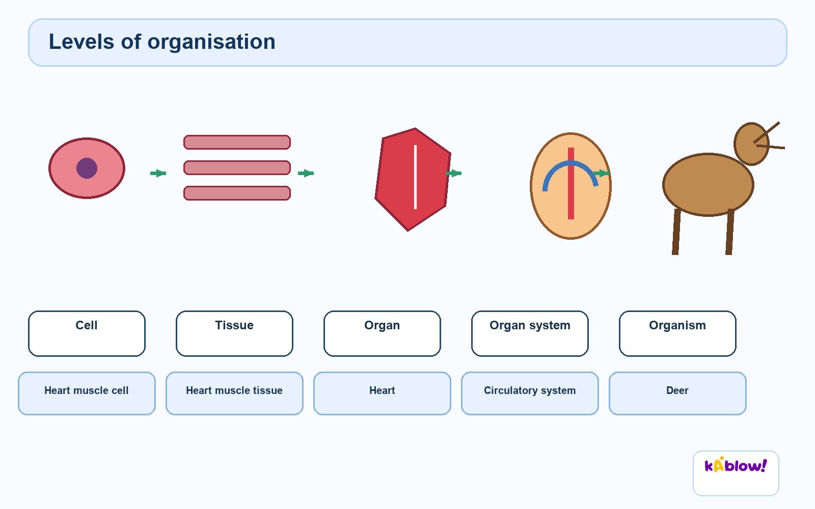
The Stomach as an Organ
- The stomach churns food to break it into smaller pieces (mechanical digestion).
- The stomach produces protease enzymes, such as pepsin, which digest proteins into amino acids.
- The stomach produces stomach acid (hydrochloric acid) which provides a suitable pH for enzymes and destroys pathogens.
- The stomach contains glandular tissue (secretes enzymes, acid, mucus), muscle tissue (contracts to churn food) and epithelial tissue (lines the stomach).
- The stomach is an example of an organ – a group of tissues working together to carry out part of digestion.
- The stomach works with other organs in the digestive system to digest food.
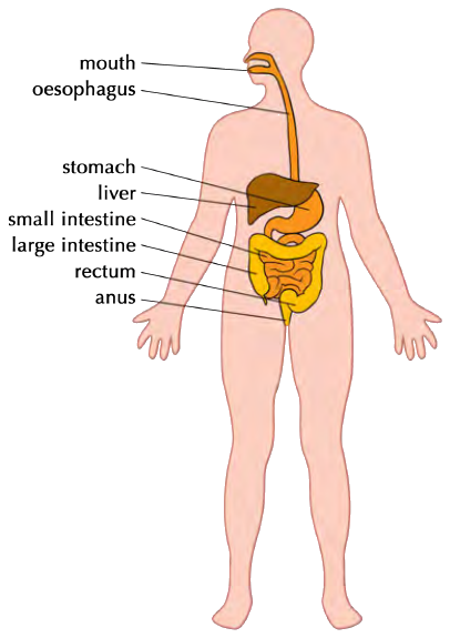
The Human Digestive System
- Digestion breaks down large, insoluble molecules into smaller, soluble molecules that can be absorbed into the bloodstream.
- Mouth: teeth break food apart (mechanical digestion); salivary glands secrete amylase which begins starch digestion into maltose.
- Oesophagus: contractions of its walls force food downwards – this is peristalsis.
- Stomach: churning continues mechanical digestion; protease enzymes begin protein digestion; hydrochloric acid provides suitable pH and kills pathogens.
- Liver: produces bile, which aids fat digestion and neutralises stomach acid. Gall bladder: stores bile before release into the duodenum via the bile duct.
- Pancreas: produces amylase, protease and lipase enzymes released into the small intestine.
- Small intestine: acidic contents neutralised by bile to slightly alkaline; enzymes complete chemical digestion; food and water absorbed into blood via villi.
- Large intestine: remaining water absorbed; solid waste forms faeces. Rectum: stores faeces. Anus: faeces leave the body (egestion).
The human digestive system with key organs labeled
Enzymes and Metabolism
- Metabolism is the sum of all reactions in a cell or organism, in which molecules are synthesised or broken down.
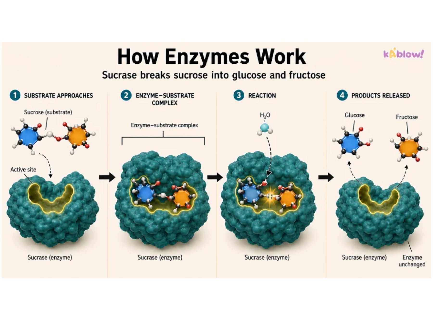
- Enzymes are biological catalysts made from protein; they speed up chemical reactions without being used up or changed.
- Enzymes allow reactions to occur at much faster speeds at relatively low temperatures, such as human body temperature.
- Substrates temporarily bind to the active site of an enzyme, forming an enzyme-substrate complex, leading to product formation.
- Enzymes are specific – usually one enzyme catalyses one particular reaction, because the active site is complementary in shape to its substrate.
- The order of amino acids determines the three-dimensional shape of an enzyme, including the shape of the active site.
The Lock and Key Model
- The 'lock and key theory' is a simplified model explaining enzyme action.
- The enzyme is like a lock, and the substrate is the key that fits perfectly into the active site.
- Step 1: Enzymes and substrates move about randomly in solution.
- Step 2: When an enzyme and its complementary substrate randomly collide, an enzyme-substrate complex forms and the reaction occurs.
- Step 3: Products form and are released from the active site; the enzyme is unchanged and can catalyse further reactions.
- For example, catalase binds to its substrate hydrogen peroxide as they are complementary in shape, whereas DNA polymerase does not.
The lock and key model
Factors Affecting Enzyme Activity: Temperature
- Enzymes work fastest at their optimum temperature – in the human body this is around 37 °C.
- Increasing temperature from 0 °C to the optimum increases enzyme activity because molecules have more kinetic energy, move faster and collide more frequently with substrate molecules.
- Low temperatures do not denature enzymes; they just make them work more slowly due to less kinetic energy.
- Heating beyond the optimum breaks the bonds holding the enzyme together; the enzyme denatures and the active site loses its shape.
- Denaturation is irreversible – substrates cannot fit into denatured enzymes and the reaction stops.
Factors Affecting Enzyme Activity: pH
- The optimum pH for most human enzymes is pH 7.
- Enzymes produced in acidic conditions, such as the stomach, have a lower optimum pH (around pH 2).
- Enzymes produced in alkaline conditions, such as the duodenum, have a higher optimum pH (around pH 8 or 9).
- If pH is too far above or below the optimum, bonds holding the amino acid chain together can be disrupted or broken.
- This changes the shape of the active site, so the substrate can no longer fit, reducing the rate of activity.
- Moving too far from the optimum pH causes the enzyme to denature and the reaction it catalyses will stop.
Enzymes and Digestion
- There are three main types of digestive enzymes: carbohydrases, proteases and lipases.
- Carbohydrases break down carbohydrates to simple sugars. Amylase breaks down starch into maltose, which is then broken down into glucose by maltase.
- Amylase is made in the salivary glands, the pancreas and the small intestine.
- Proteases break down proteins into amino acids in the stomach and small intestine; they are made in the stomach (pepsin), pancreas and small intestine.
- Lipases break down lipids (fats) into glycerol and fatty acids; they are produced in the pancreas and secreted into the duodenum.
- Bile is alkaline and neutralises hydrochloric acid from the stomach, providing the alkaline optimum pH for small intestine enzymes.
- Bile also emulsifies large drops of fat into smaller ones, increasing surface area so lipase breaks down fat faster.
Required Practical: Enzymes and pH
- Aim: to investigate the effect of pH on the rate of reaction of amylase.
- Amylase digests starch (a polysaccharide of glucose) into maltose (a disaccharide of glucose).
- Iodine solution is used as an indicator: it turns blue-black in the presence of starch and remains orange-brown when starch is absent.
- Method: place drops of iodine on a spotting tile; mix amylase, pH buffer and starch solution; sample the mixture every 10 seconds onto the iodine.
- Repeat until the iodine remains orange-brown – this means all starch has been digested.
- The less time the iodine takes to remain orange-brown, the faster the enzyme works at that pH.
- Control temperature using a water bath at 35 °C; allow all solutions to reach the temperature before use.
- A colorimeter can measure the progress of the reaction more accurately by measuring absorbance or transmission of light.
Required Practical: Food Tests
- Aim: to use qualitative reagents to test for carbohydrates, lipids and proteins in foods.
- Iodine test for starch: orange-brown reagent turns blue-black if starch is present.
- Benedict's test for sugar: light blue reagent turns green to brick-red if sugar is present; requires heating in a water bath.
- Ethanol emulsion test for lipids: colourless reagent turns cloudy if lipids are present.
- Biuret test for protein: blue reagent turns lilac-purple if protein is present.
- Prepare solid food samples by breaking them up with a pestle and mortar, adding distilled water, stirring and filtering.
- Hazards: wear goggles (Biuret contains copper(II) sulfate; iodine is an irritant); sodium hydroxide in Biuret is corrosive; ethanol is highly flammable and must be kept away from the Bunsen burner.
Diapositivas
Sign up free to view the lesson slides
Step through every slide for this topic — plus flashcards and revision notes — with a free account.
Preguntas de práctica
Vista previa gratis — 8 de 58 preguntas. Regístrate para verlas todas.
1.Which term describes the hydrogen peroxide in the reaction catalysed by catalase?
Easy- Aproduct
- Bsubstrate
- Cactive site
- Dcontrol
2.Potato cells contain the enzyme catalase. This enzyme catalyses the breakdown of hydrogen peroxide into oxygen and water. Which of the following is the substrate in this reaction?
Easy- Aoxygen
- Bwater
- Chydrogen peroxide
- Dcatalase
3.Which of the following is the best definition of an enzyme?
Easy- AA biological catalyst made from protein
- BA biological catalyst made from carbohydrate
- CA biological catalyst made from lipid
- DA biological catalyst made from nucleic acid
4.Which of the following is the main function of the diaphragm?
Easy- AIt helps change the volume of the thorax to allow inhalation and exhalation
- BIt produces mucus to trap dust
- CIt transports oxygen into the blood
- DIt protects the lungs from damage
5.Which of the following structures is the windpipe that connects the mouth and nose to the lungs?
Easy- ABronchus
- BBronchiole
- CTrachea
- DAlveolus
6.Which of the following is NOT a feature of gas exchange surfaces?
Medium- ALarge surface area
- BThin walls
- CGood blood supply
- DThick walls to increase diffusion distance
7.Which of the following is the correct pathway of blood through the heart?
Easy- AVena cava → right atrium → right ventricle → pulmonary artery → lungs
- BVena cava → left atrium → left ventricle → pulmonary vein → lungs
- CPulmonary vein → right atrium → right ventricle → aorta → lungs
- DAorta → left atrium → left ventricle → pulmonary artery → lungs
8.Which of the following blood vessels carries oxygenated blood away from the heart?
Easy- APulmonary artery
- BPulmonary vein
- CVena cava
- DAorta
Unlock all 58 questions, flashcards & more
Crea una cuenta gratis para ver todas las preguntas, las diapositivas, las tarjetas y los apuntes de repaso de este tema.