Cell & Nuclear Division
खेलकर सीखें
इन सवालों के जवाब देकर एनर्जी कमाएं, फिर मछली पकड़ें और घूमें। कोई अकाउंट नहीं चाहिए।
लेसन नोट्स
Cell Division and Cytokinesis
- According to cell theory, new cells are produced from pre-existing ones; a parent cell divides to produce two daughter cells.
- Nuclear division occurs first, followed by cytokinesis, the division of the cytoplasm, which produces two genetically identical daughter cells in mitosis.
- In animal cells, cytokinesis involves a cleavage furrow formed by a contractile ring of actin and myosin proteins that pinches the cell in two.
- In plant cells, a cell plate forms at the equator and develops into a new cell wall, separating the daughter cells.
- Cytokinesis is usually equal, but can be unequal, as in oogenesis (forming an ovum and polar bodies) and budding in yeast.
- Each daughter cell must receive at least one mitochondrion (and a chloroplast in plant cells) because these organelles can only be made by dividing pre-existing structures.
Nuclear Division: Mitosis and Meiosis
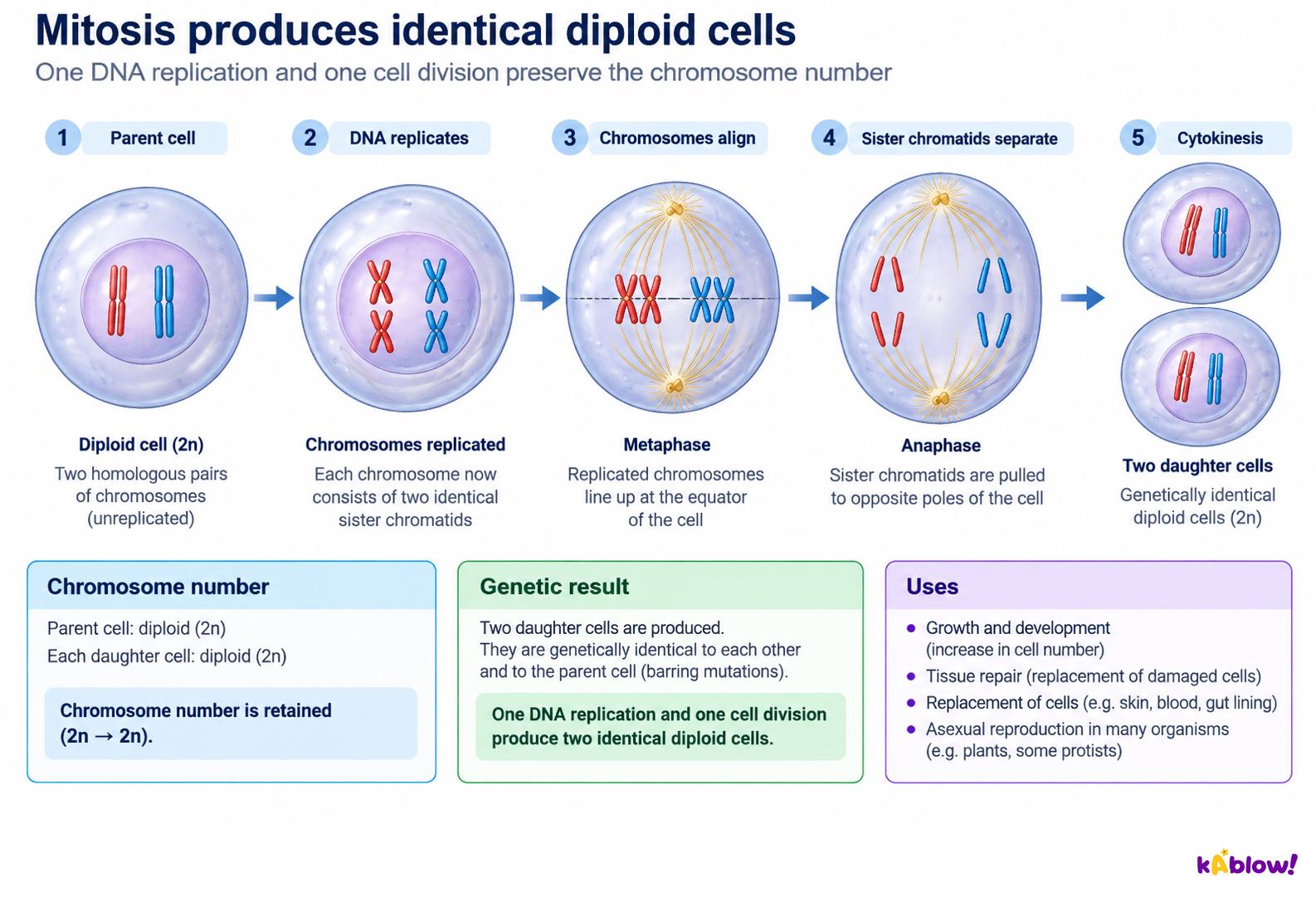
- Mitosis produces two genetically identical daughter nuclei that are also identical to the parent nucleus, maintaining the chromosome number and genome.
- Mitosis is used for growth, repair of damaged tissues, replacement of cells, and asexual reproduction.
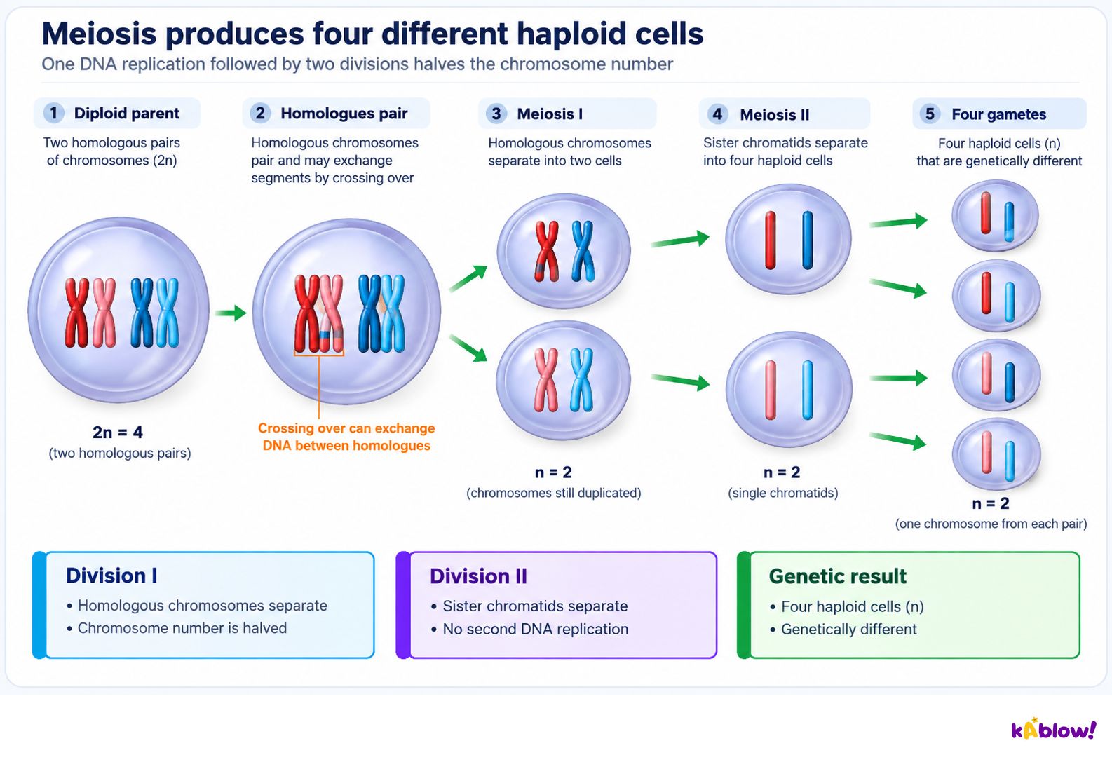
- Meiosis produces four genetically different daughter cells, each with half the number of chromosomes of the parent cell (haploid, n).
- Meiosis is used to produce gametes (sex cells) and is important for generating genetic diversity.
- Before nuclear division, DNA replication occurs during interphase, creating two identical sister chromatids joined at a centromere.
- During anaphase, sister chromatids separate and are pulled to opposite poles; after separation, each chromatid is considered an individual chromosome.
Meiosis
Chromosome Structure and Condensation
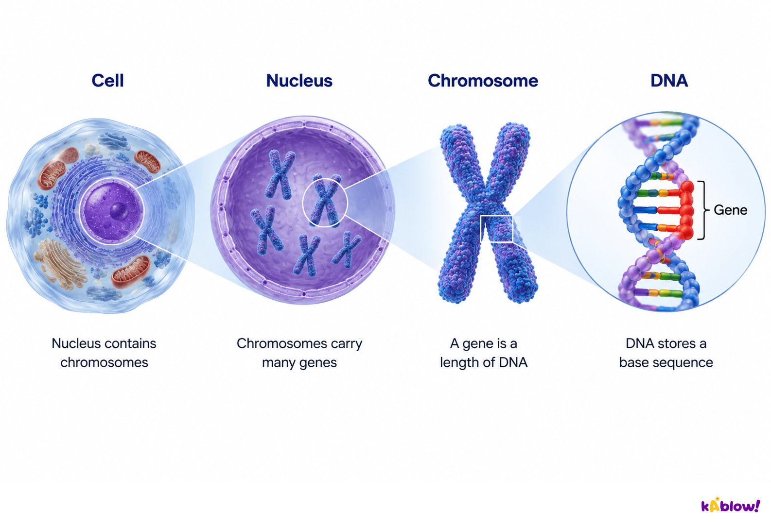
- DNA is a very long molecule that must fit inside a much smaller nucleus; it is packaged with histone proteins.
- DNA wraps around eight histone proteins to form a nucleosome; the complex of DNA and histones is called chromatin.
- During prophase, chromatin condenses by supercoiling to form visible chromosomes.
- Microtubules are tubulin fibres that form part of the cytoskeleton and are responsible for chromosome movement during cell division.
- Microtubules are made of α-tubulin and β-tubulin dimers that can be added or removed to change length.
- Motor proteins carry chromosomes along microtubules to the equator of the cell.
From cell to gene
Phases of Mitosis
- Prophase: chromosomes condense and become visible; centrosomes move to opposite poles; spindle fibres emerge; the nuclear envelope breaks down and the nucleolus disappears.
- Metaphase: chromosomes line up at the equator (metaphase plate); spindle fibres attach to centromeres via kinetochores; each sister chromatid is attached to a spindle fibre from opposite poles.
- Anaphase: sister chromatids separate at the centromere; spindle fibres shorten and pull the separated chromosomes to opposite poles.
- Telophase: chromosomes arrive at poles and decondense; nuclear envelopes reform; spindle fibres break down; new nucleoli form.
- Cytokinesis follows nuclear division, dividing the cytoplasm to form two daughter cells.
- Remember the order of mitosis stages with PMAT: Prophase, Metaphase, Anaphase, Telophase.
Mitosis
Identifying Mitosis Stages in Micrographs
- Interphase: most cells are in this stage; chromatin is visible but chromosomes are not, so nuclei appear dark.
- Prophase: chromosomes are visible and the nuclear envelope is breaking down; only one group of chromosomes is present.
- Metaphase: chromosomes are lined up along the middle of the cell.
- Anaphase: chromosomes are moving away from the middle towards opposite poles, often appearing V-shaped.
- Telophase: chromosomes have arrived at opposite poles, begin to uncoil, and the nuclear envelope is reforming; two groups of chromosomes are present.
- Cytokinesis: in animal cells a cleavage furrow forms; in plant cells a cell plate forms at the metaphase plate and expands towards the cell wall.
Meiosis and Genetic Variation
- Meiosis involves two divisions: Meiosis I (reduction division) separates homologous chromosomes, and Meiosis II separates sister chromatids.
- During prophase I, homologous chromosomes pair up to form a bivalent; this pairing is called synapsis.
- Crossing over occurs between non-sister chromatids at points called chiasmata, exchanging genetic material and producing new allele combinations.
- Random orientation of bivalents at metaphase I means each homologous chromosome is randomly attached to one pole, and bivalents assort independently.
- The number of possible chromosome combinations from random assortment is 2n, where n is the number of homologous pairs.
- Non-disjunction occurs when chromosomes fail to separate correctly during anaphase I or II, leading to gametes with an abnormal chromosome number.
- Down syndrome (Trisomy 21) is caused by non-disjunction of chromosome 21, resulting in three copies and a total of 47 chromosomes.
The Cell Cycle and Its Control
- The cell cycle is the regulated sequence of events between one cell division and the next, consisting of interphase, nuclear division (mitosis), and cell division (cytokinesis).
- Interphase includes G1, S, and G2 phases; it is the longest and most active phase, where the cell grows and carries out normal functions.
- G1 phase: cell grows, synthesises RNA, enzymes, and proteins; may receive signal to divide.
- S phase: DNA replication occurs, resulting in chromosomes with two identical sister chromatids.
- G2 phase: cell continues to grow, DNA is checked and repaired, and proteins for division (e.g., tubulin) are produced.
- The cell cycle is controlled by cyclins (proteins) and cyclin-dependent kinases (enzymes); cyclins bind to kinases to trigger specific events.
- There are three checkpoints: at G1, G2, and M; these must be overridden before the next stage begins.
Uncontrolled Cell Division and Tumour Formation
- Mutations in genes controlling the cell cycle can lead to cancer.
- Proto-oncogenes code for proteins that stimulate normal cell division; mutation converts them to oncogenes, leading to increased or permanently activated proteins and uncontrolled division.
- Tumour-suppressor genes code for proteins that inhibit cell division or promote apoptosis; mutations may result in reduced or deactivated proteins, also leading to uncontrolled division.
- Malignant tumours are cancerous: they grow rapidly, invade and destroy surrounding tissues, and can metastasise (spread via blood or lymph).
- Benign tumours are not cancerous: they grow slowly, do not invade other tissues, and do not metastasise.
- Carcinogens such as UV radiation, tobacco, asbestos, and processed meat can initiate malignant tumour formation.
Mitotic Index
- The mitotic index is the proportion of cells in a sample that are undergoing mitosis.
- It is calculated using the formula: mitotic index = number of cells with visible chromosomes / total number of cells.
- The answer can be multiplied by 100 to give a percentage.
- A high mitotic index indicates a high rate of cell division, which may suggest the presence of a tumour.
- To study mitosis, root tip meristems can be used; cells are stained and squashed to spread them out for observation.
- The mitotic index formula must be remembered as it is not provided in exams.
स्लाइड्स
Sign up free to view the lesson slides
Step through every slide for this topic — plus flashcards and revision notes — with a free account.
प्रैक्टिस सवाल
फ्री प्रीव्यू — 64 में से 8 सवाल। सभी देखने के लिए साइन अप करें।
1.Which of the following is the correct definition of the mitotic index?
Easy- AThe proportion of cells in a sample that are undergoing mitosis
- BThe number of cells in a sample that are in interphase
- CThe total number of cells in a sample
- DThe number of cells that are undergoing meiosis
2.Which of the following is the correct formula for calculating the mitotic index?
Easy- Anumber of cells in interphase / total number of cells
- Bnumber of cells undergoing mitosis / total number of cells
- Ctotal number of cells / number of cells undergoing mitosis
- Dnumber of cells undergoing mitosis / number of cells in interphase
3.Mitosis produces two genetically identical daughter nuclei.
EasyTrue or false?
4.Meiosis produces four haploid nuclei from one diploid nucleus.
EasyTrue or false?
5.Which of the following correctly describes the term 'chromatid'?
Easy- AA single strand of DNA that is replicated during interphase
- BOne of the two identical strands of DNA that make up a chromosome after DNA replication
- CThe region where two chromatids are joined together
- DA pair of homologous chromosomes
6.What is the name of the narrow region that joins two sister chromatids together?
Easy- ACentromere
- BCentrosome
- CChiasma
- DKinetochore
7.Which of the following statements about homologous chromosomes is correct?
Easy- AThey carry the same genes in the same order but may have different alleles
- BThey are identical copies of each other produced by DNA replication
- CThey are only found in haploid cells
- DThey are separated during anaphase of mitosis
8.Crossing over occurs between non-sister chromatids of homologous chromosomes.
EasyTrue or false?
Unlock all 64 questions & more
इस टॉपिक के हर सवाल, स्लाइड्स, फ्लैशकार्ड और रिवीज़न नोट्स देखने के लिए फ्री अकाउंट बनाएं।