Nucleic Acids
खेलकर सीखें
इन सवालों के जवाब देकर एनर्जी कमाएं, फिर मछली पकड़ें और घूमें। कोई अकाउंट नहीं चाहिए।
लेसन नोट्स
DNA and RNA as Genetic Material
- DNA (deoxyribonucleic acid) carries the genetic code in all living organisms, making the genetic code universal.
- DNA is mainly found in the nucleus where it forms chromosomes, and is also found in chloroplasts and mitochondria of eukaryotic cells.
- RNA (ribonucleic acid) is the main component of ribosomes and plays an important role in protein synthesis; some RNA is also found in the nucleus and cytoplasm.
- Certain viruses (such as SARS-CoV-2) contain RNA as their genetic material instead of DNA, causing diseases such as COVID-19, Ebola, mumps and influenza.
- Viruses are not considered living because they cannot replicate by themselves, depend on other living cells, and lack a cellular structure.
From cell to gene
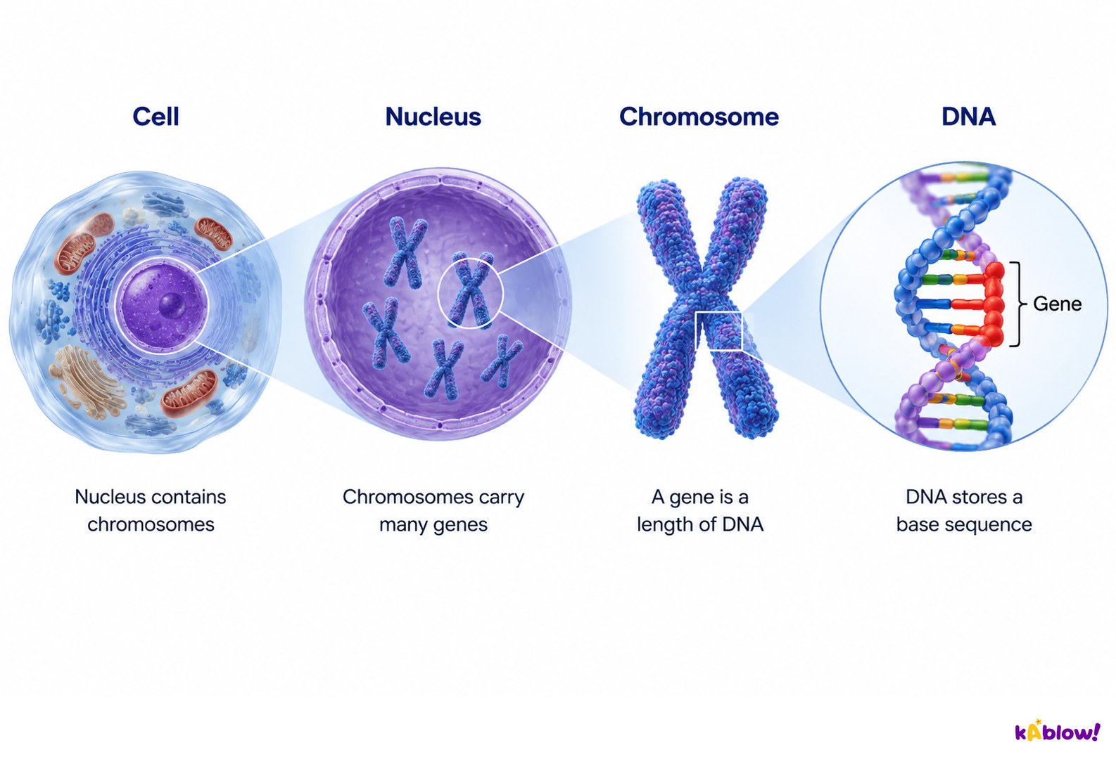
Nucleotide Components
- Both DNA and RNA are polymers made of repeating units called nucleotides.
- Each nucleotide is formed from a pentose sugar (5 carbon atoms), a nitrogen-containing organic base (1 or 2 rings), and a phosphate group (acidic and negatively charged).
- The base and phosphate group are both covalently bonded to the sugar.
- The nitrogenous bases in DNA are adenine (A), guanine (G), cytosine (C) and thymine (T); RNA shares the same bases except thymine is replaced by uracil (U).
- Adenine and guanine are purine bases; cytosine, thymine and uracil are pyrimidine bases.
The sugar-phosphate backbone
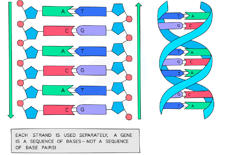
Drawing Nucleotides and Polynucleotides
- Simple shapes can represent nucleotide components: pentagons for pentose sugars, circles for phosphates (often with P inside), and rectangles for bases.
- Covalent bonds can be shown with solid lines; hydrogen bonds with dashed lines or complementary shapes that fit together.
- Nucleotides join in chains when the phosphate group of one nucleotide forms a covalent bond to the pentose sugar of the next, forming a large polymer.
- These polymers are called nucleic acids or polynucleotides; the linkage forms a sugar-phosphate backbone with a base linked to each sugar.
- The polymer of nucleotides is known as a strand; DNA is double-stranded, RNA is usually single-stranded.
- There are just 4 separate bases that can be joined in any combination/sequence because the sugar and phosphate are the same in every nucleotide.
RNA Structure
- RNA molecules are relatively short (a hundred to a few thousand nucleotides) and usually form a single-stranded polynucleotide with ribose as the pentose sugar.
- RNA nucleotides contain adenine, guanine, cytosine and uracil (instead of thymine).
- The carbon atoms in nucleotides are numbered from the right in a clockwise direction, indicating the orientation of the polynucleotide.
- Different types of RNA: mRNA (formed in the nucleus, transported to ribosomes), tRNA (transports amino acids to ribosomes), and rRNA (forms part of ribosomes).
- Adjacent RNA nucleotides are linked by condensation reactions, releasing a molecule of water and forming a phosphodiester bond between the pentose sugar of one nucleotide and the phosphate group of the next.
DNA Structure and Complementary Base Pairing
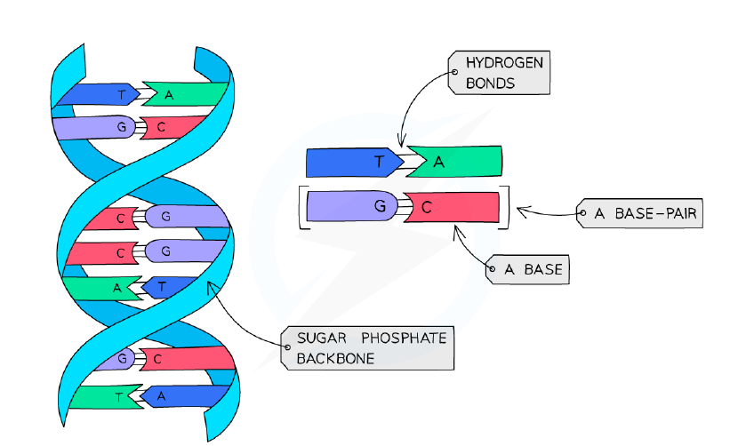
- DNA is a double helix made of two antiparallel strands of nucleotides linked by hydrogen bonding between complementary base pairs.
- Each DNA polynucleotide strand has alternating deoxyribose sugars and phosphate groups forming the sugar-phosphate backbone, with a 3' end and a 5' end.
- Because the strands run in opposite directions, one is the 5' to 3' strand and the other is the 3' to 5' strand.
- The nitrogenous bases project out from the backbone towards the interior of the double-stranded DNA molecule.
- Adenine (A) always pairs with thymine (T) via two hydrogen bonds; guanine (G) always pairs with cytosine (C) via three hydrogen bonds — this is complementary base pairing.
- The base pairings A–T and C–G are equal in length, so the DNA helix has the same 3D structure regardless of base sequence; purines are larger than pyrimidines due to their two carbon ringed structure.
The DNA double helix
Basis of the Genetic Code
- DNA molecules carry the genetic code as a sequence of nitrogenous bases (A, G, C, T) in the nucleotides.
- One strand carries the base sequence read by enzymes — the coding strand; the sequence of bases forming genes determines the order of amino acids in synthesised proteins.
- The code is read as a triplet of bases called a codon, with each sequence of three bases coding for one amino acid; there are 20 different amino acids.
- The sequence of amino acids determines the shape and function of the protein synthesised from the code.
- The genetic code is universal — almost every organism uses the same code (with rare minor exceptions), so the same triplet codes code for the same amino acids in all living things.
- The universal nature of the genetic code is why genetic engineering is possible and provides evidence for a universal common ancestor.
- Conserved sequences are coding and non-coding sequences that have remained unchanged in all organisms; highly conserved sequences are usually found in genes for proteins involved in transcription and translation, and in histone proteins.
DNA vs RNA Comparison
- Pentose sugar: DNA contains deoxyribose; RNA contains ribose.
- Bases: DNA contains A, C, G, T; RNA contains A, C, G, U (uracil instead of thymine).
- Number of strands: DNA is double-stranded (double helix); RNA is single-stranded.
- RNA polynucleotide chains are relatively short compared to DNA.
- Complementary base pairing means the base sequence on one DNA strand determines the sequence of the other — one strand acts as a template of the other.
- This allows DNA to be copied very precisely during DNA replication, ensuring the genetic code is accurately copied and expressed in newly formed cells.
DNA as an Information Storage Molecule
- Despite only four bases (A, T, C, G), they can combine to form a very diverse range of DNA base sequences in molecules of different lengths.
- DNA therefore has an almost limitless capacity for storing genetic information.
- Storage capacity can be measured by the number of genes; even the most simplistic forms of life may contain several thousand genes.
- Examples of approximate gene numbers: Human 20 000, Dog 19 000, Water flea 31 000, Bacterium (E. coli) 4 300, Rice plant 41 500.
- Storage capacity can also be measured in base pairs; the DNA in a human cell nucleus contains about 3.2 gigabases (about 10⁹ DNA base pairs).
- These base pairs are contained in DNA with a length of about 2 meters, fitting within the nucleus of each human cell — an indication of how incredibly well packaged this genetic information is.
Nucleosomes and Molecular Visualisation Software
- Unlike most prokaryotic DNA which is 'naked', eukaryotic nuclear DNA is associated with proteins called histones to form chromatin.
- A nucleosome consists of a strand of DNA coiled around a core of eight histone proteins (octamer) to form a bead-like structure.
- DNA takes two turns around the histone core and is held in place by an additional histone protein attached to linker DNA; the DNA continues to wind around a series of nucleosomes forming a 'string of beads'.
- Nucleosomes help to supercoil the DNA, resulting in a compact structure that saves space within the nucleus; they also help to protect DNA and facilitate movement of chromosomes during cell division.
- Nucleosomes can be tagged with proteins to promote or suppress transcription.
- Molecular visualisation software allows macromolecules (protein, DNA, RNA, complex carbohydrates) to be visualised as 3-D structures, relating primary sequence information to structure and function.
- Macromolecules can be represented as ball and stick atom models or simplified ribbon representations; most software is freely available online or through bioinformatics repositories such as the Protein Data Bank (PDB).
- In the PDB, searching for 6T79 (human nucleosome) and selecting '3D view' in Mol×shows the DNA double helix surrounding the histone proteins, with the DNA making two loops around the histone octamer core and histone tails projecting from the core.
The Hershey & Chase Experiment
- DNA was identified in 1869 but many scientists assumed protein was the heritable material, owing to there being 20 amino acids and only 4 nucleotide bases.
- In the 1950s, Alfred Hershey and Martha Chase showed that DNA, not protein, is the factor of heredity responsible for carrying genetic information from one generation to another.
- They used viruses that infect bacteria (T2 bacteriophage infecting E. coli) as they consist only of DNA encapsulated by a protein coat.
- Their method relied on a chemical difference: DNA contains phosphorus but no sulfur, while proteins contain sulfur but no phosphorus, allowing a radioactive isotope of each element to label one biomolecule without labelling the other.
- Step 1: Bacteria were grown in media containing either radioactive sulfur (³⁵S) or radioactive phosphorus (³²P), then infected with viruses; progeny viruses took up the radioactive atoms, giving ³⁵S-labelled protein coats or ³²P-labelled DNA.
- Step 2: Fresh, unlabelled bacteria were infected separately with each type of labelled virus; whichever biomolecule entered the bacteria must be the heritable material.
- Step 3: A blender detached the viral coats from the bacterial cells, and centrifugation separated them — viruses are small and remained in the supernatant; bacteria are larger and formed a pellet.
- Results: With ³²P-labelled viruses, radioactivity was found in the pellet (viral DNA had entered the bacteria); with ³⁵S-labelled viruses, radioactivity remained in the supernatant (protein coats had stayed outside).
- Conclusion: DNA, not protein, is the hereditary material — providing unequivocal proof.
- NOS: The availability of radioisotopes as research tools (made available at the end of the Second World War) made the Hershey-Chase experiment possible.
Chargaff's Data and the Problem of Induction
- Erwin Chargaff analysed the DNA composition of different organisms during the 1930s and 1940s and discovered that the number of purine bases equals the number of pyrimidine bases.
- He also found that the number of adenine bases equals the number of thymine bases, while the number of guanine bases equals the number of cytosine bases.
- This means a purine base can only pair with a pyrimidine base between the sugar-phosphate backbone, since they have different sizes — forming the foundation of complementary base pairing in DNA.
- The inductive scientific method starts with observations and raw data, then formulates a hypothesis tested by investigation; using past data to predict the future assumes the future will be the same.
- It is impossible to prove a hypothesis generated by inductive reasoning as absolutely true — this is the problem of induction, the main reason most scientific theories are considered tentative.
- Even if several investigations support a hypothesis, it can still be falsified in the future as new discoveries are made; Karl Popper suggested new scientific knowledge is gained by the falsification of existing hypotheses rather than by inductive steps.
- Phoebus Levene discovered the pentose sugars of DNA and RNA in the early 1900s and suggested the tetranucleotide hypothesis — that nucleic acid structure was a repeating tetramer unit called a nucleotide.
- At the time, limitations to analytic techniques made it difficult to determine the relative amounts of nucleotides present in nucleic acids.
- The tetranucleotide hypothesis was falsified by Chargaff's data in the late 1940s, which showed the organism-specificity of nucleic acids; when the structure of DNA was determined in the 1950s, it further proved the repeating tetramer unit would not be suitable for carrying genetic information from one generation to the next.
स्लाइड्स
Sign up free to view the lesson slides
Step through every slide for this topic — plus flashcards and revision notes — with a free account.
प्रैक्टिस सवाल
फ्री प्रीव्यू — 60 में से 8 सवाल। सभी देखने के लिए साइन अप करें।
1.Which of the following correctly lists the three components of a nucleotide?
Easy- AA pentose sugar, a nitrogen-containing base and a phosphate group
- BA hexose sugar, an amino acid and a phosphate group
- CA pentose sugar, a nitrogen-containing base and a sulfate group
- DA deoxyribose sugar, a protein and a phosphate group
2.The diagram represents a nucleotide. Which row correctly identifies components 1, 2 and 3?
Easy- A1 = Phosphate group, 2 = Pentose sugar, 3 = Adenine
- B1 = Adenine, 2 = Pentose sugar, 3 = Phosphate group
- C1 = Pentose sugar, 2 = Phosphate group, 3 = Adenine
- D1 = Phosphate group, 2 = Adenine, 3 = Pentose sugar
3.Which of the following statements apply to RNA? (select all that apply)
Medium- AThe molecule is in the shape of a double helix, with antiparallel strands
- BThe four bases are adenine, cytosine, guanine, thymine
- CRibose is present as the pentose sugar in its nucleotides
- DIt is a polymer formed by the linkage of many nucleotides
- EIt is usually single-stranded
4.The diagram shows a section of a DNA molecule. Which of these base pairs are not correct?
Medium- ANone
- B1
- C2
- D3
5.Which of the following statements correctly describes a nucleosome?
Easy- AA strand of DNA coiled around a central core of eight histone proteins.
- BA strand of DNA wrapped around a central core of four histone proteins.
- CA section of supercoiled DNA.
- DA strand of DNA wrapped around a central core of eight chromatin proteins.
6.A short piece of DNA 19 base pairs long was analysed to find the number of nucleotide bases in each of the polynucleotide strands. Some of the results are shown below. Number of nucleotide bases A T G C Strand 1 8 - - - Strand 2 - 8 3 4 How many nucleotides containing adenine (A) were present in strand 2?
Medium- A2
- B4
- C6
- D8
7.Which of the following would be a result of analysing a DNA molecule?
Medium- AAn equal ratio of pentose sugars to phosphates and an equal ratio of thymine to adenine bases.
- BTwice as many pentose sugars as phosphates and an equal ratio of guanine to cytosine bases.
- CAn equal ratio of hexose sugars to phosphates and an equal ratio of thymine to cytosine bases.
- DTwice as many hexose sugars as phosphates and an equal ratio of guanine to adenine bases.
8.DNA is described as a double-helix, antiparallel structure. Which of the following statements best explains the term 'antiparallel'?
Medium- AOne strand possesses deoxyribose sugar, the opposite strand possesses ribose sugars.
- BThe nitrogenous bases invert their orientation on the sense strand versus the antisense strand.
- CThe alignment of the sugar-phosphate backbone of each strand runs in opposite directions.
- DThe alignment of the two strands of nucleotides runs in opposite directions.
Unlock all 60 questions & more
इस टॉपिक के हर सवाल, स्लाइड्स, फ्लैशकार्ड और रिवीज़न नोट्स देखने के लिए फ्री अकाउंट बनाएं।