Cell structure
遊んで学ぼう
問題に答えてエネルギーを集めたら、釣りや探検を楽しもう。アカウント不要。
レッスンノート
Eukaryotic and Prokaryotic Cells
- Eukaryotic cells have their genetic material (DNA) enclosed within a nucleus; they include animal, plant, fungal and protoctist cells.
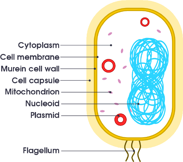
- Prokaryotic cells (e.g. bacteria) do not have a nucleus; their DNA is a single loop in the cytoplasm, and they may also contain plasmids (small rings of DNA).
- All cells have a cell membrane, cytoplasm and ribosomes; prokaryotic cells also have a cell wall, but it is made of peptidoglycan, not cellulose.
- Prokaryotic cells are much smaller than eukaryotic cells, typically about 1 μm compared to 10–100 μm for eukaryotic cells.
- Some bacteria have a flagellum (a tail-like structure) that allows them to move.
- Plasmids contain extra genes not found in the main chromosomal DNA.
Diagram of a prokaryotic cell with labeled parts.
Animal and Plant Cells
- Animal cells contain a nucleus, cytoplasm, cell membrane, mitochondria and ribosomes.
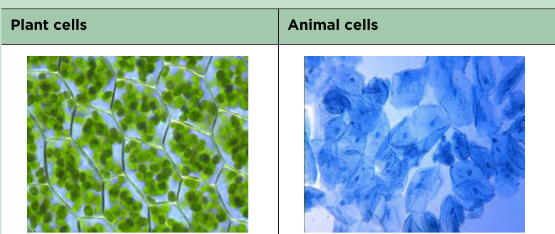
- Plant cells have all the structures found in animal cells, plus a cell wall made of cellulose, a permanent vacuole filled with cell sap, and (in leaf and stem cells) chloroplasts.
- The nucleus contains genetic material and controls the cell's activities.
- Mitochondria are the site of aerobic respiration, releasing energy for the cell.
- Ribosomes are the site of protein synthesis.
- Chloroplasts contain chlorophyll and carry out photosynthesis.
- The cell wall provides support and defines the cell's shape; the cell membrane controls what enters and leaves the cell.
Plant cells (left), packed with green chloroplasts, next to animal cells (right), which have no chloroplasts or cell wall.
Cell Specialisation
- Specialised cells have adaptations (in shape or sub-cellular structures) that help them carry out a particular function.
- Sperm cells: the tail allows movement, the mid-piece has many mitochondria to release energy, and the head contains enzymes to digest the egg's outer layer.
- Nerve cells: elongated to conduct electrical impulses over long distances; dendrites connect to other cells.
- Muscle cells: contain protein filaments that slide to allow contraction, and many mitochondria to release energy.
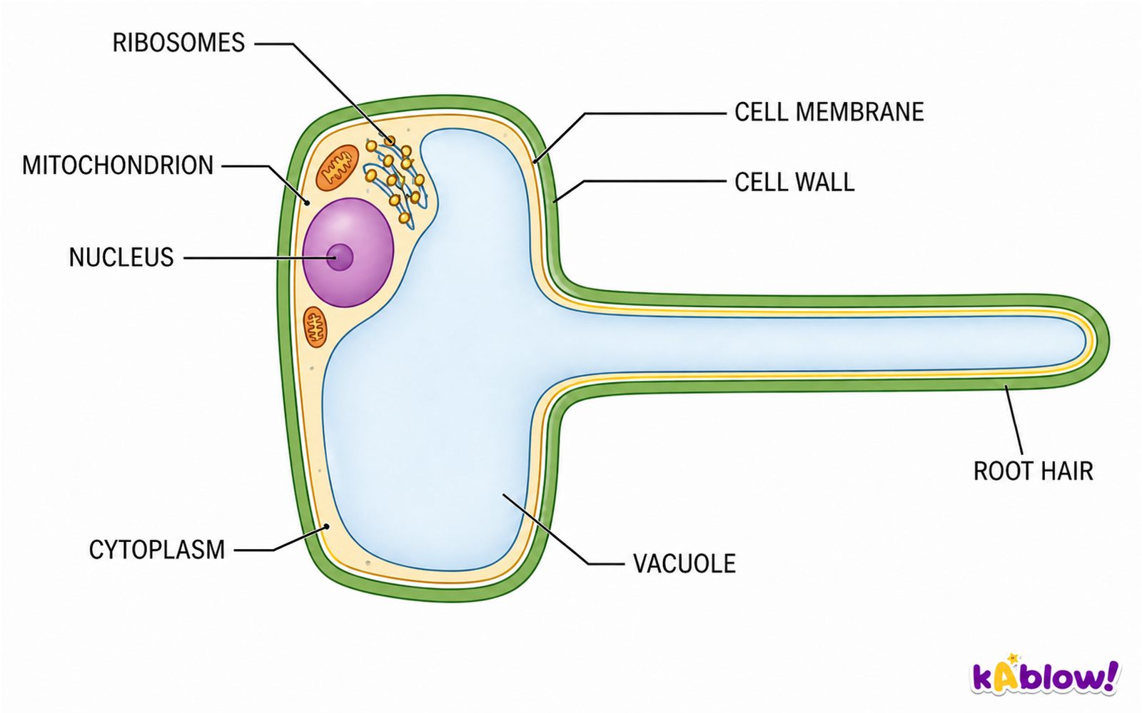
- Root hair cells: have an extension that increases surface area for water absorption, and mitochondria for active transport of mineral ions.
- Xylem cells: dead, hollow tubes with no end walls, strengthened by lignin, for transporting water and minerals.
- Phloem cells: living cells with sieve plates, for transporting dissolved sugars and amino acids.
Root hair cell
Cell Differentiation
- Differentiation is the process by which a cell changes to become specialised, developing the structure and sub-cellular components needed for its function.
- Almost all cells in a multicellular organism contain the same genes, but only some genes are switched on in each cell type.
- In animals, most cells differentiate early in development; mature animals mainly use cell division for repair and replacement.
- Some animal cells (adult stem cells) retain the ability to differentiate throughout life.
- Plant cells retain the ability to differentiate throughout the plant's life, in regions called the meristem.
Microscopy
- Light microscopes use light and lenses to magnify specimens; they can show cells and large structures like nuclei, often with the help of stains.
- Electron microscopes use beams of electrons, giving much higher magnification and resolution, so they reveal smaller structures such as mitochondria and ribosomes.
- Magnification = image size ÷ actual size. Rearranged: actual size = image size ÷ magnification; image size = magnification × actual size.
- Total magnification of a light microscope = eyepiece lens magnification × objective lens magnification.
- Magnification has no units and is written as, for example, ×100.
- Resolution is the ability to distinguish two points that are close together; it is not the same as magnification.
Required Practical: Using a Light Microscope
- Prepare a slide of animal cells (e.g. cheek cells) or plant cells (e.g. onion cells).
- For a solid specimen: cut a thin layer of tissue, place it on the slide, add a stain if needed (e.g. iodine for onion, methylene blue for cheek cells), then lower a coverslip to avoid air bubbles.
- For a liquid specimen: add a drop to the slide, cover with a coverslip, and press gently to remove air bubbles.
- Always start with the low-power objective lens to find the specimen and avoid damaging the slide or lens.
- When drawing, use a sharp pencil, continuous lines, no shading, and include a magnification or scale bar.
- Add a drop of water to the specimen to prevent dehydration, and take care with sharp instruments and stains.
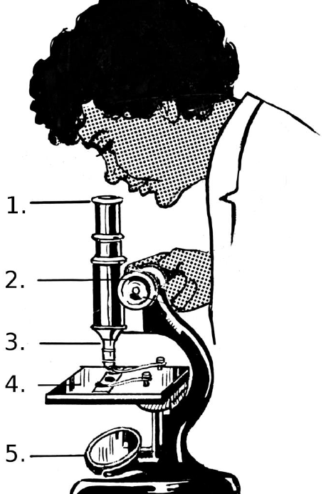
Parts of a light microscope
Culturing Microorganisms
- Bacteria can be grown in a nutrient broth or on an agar gel plate.
- Aseptic technique is used to avoid contamination: sterilise Petri dishes and media before use, and pass the inoculating loop through a flame before transferring bacteria.
- After adding bacteria, secure the lid with adhesive tape and store the plate upside down.
- In school laboratories, cultures should be incubated at a maximum of 25 °C to avoid growing harmful human pathogens.
- Bacteria divide by binary fission; under ideal conditions they can divide as often as every 20 minutes.
- To calculate the number of bacteria after a given time, use the mean division time and express the answer in standard form if required.
Required Practical: Investigating Antibiotics and Antiseptics
- Place paper discs soaked in different antibiotics or antiseptics on an agar plate that has been inoculated with bacteria.
- Incubate the plate for a set time, then measure the zone of inhibition (clear area) around each disc.
- The area of a clear zone is calculated using the formula: area = πr², where r is the radius (half the diameter).
- The larger the zone of inhibition, the more effective the substance is at killing bacteria.
- Control variables include the concentration of the substance, the size of the disc, and the incubation temperature and time.
- To make results valid, use aseptic technique, repeat the investigation, and calculate a mean.
Units and Standard Form
- Cells are measured in micrometres (μm): 1 mm = 1000 μm, 1 μm = 1000 nm.
- Convert units before doing magnification calculations: e.g. 1 μm = 0.001 mm = 1 × 10⁻³ mm.
- Standard form is written as a × 10ⁿ, where 1 ≤ a < 10.
- For large numbers, n is positive; for small numbers, n is negative.
- Order of magnitude is a difference of a factor of 10; e.g. a 10 μm cell is one order of magnitude larger than a 1 μm cell.
スライド
練習問題
無料プレビュー — 60問中8問。すべて見るには登録を。
1.Which of the following structures is not found in a prokaryotic cell?
Easy- ACellulose cell wall
- BRibosomes
- CPlasmids
- DA circular loop of DNA
2.Bacteria reproduce by a process called binary fission. Which of the following best describes binary fission?
Easy- AA cell divides to produce four daughter cells
- BA cell divides to produce two identical cells
- CTwo cells join together to form one larger cell
- DA cell produces a copy by budding
3.One bacterium divides five times. How many bacteria are present in the population now?
Easy- A6
- B32
- C16
- D64
4.A prokaryotic cell contains its genetic material enclosed within a nucleus.
EasyTrue or false?
5.Plant cells do not need mitochondria because they can carry out photosynthesis.
EasyTrue or false?
6.Which sub-cellular structure controls the activities of the cell?
Easy- AThe cell membrane
- BThe cytoplasm
- CThe nucleus
- DThe ribosome
7.A student says the cell wall controls which substances enter and leave a plant cell. Which statement corrects this misconception?
Medium- AThe cell wall is the site of protein synthesis
- BThe cell membrane controls what enters and leaves the cell
- CThe vacuole controls what enters and leaves the cell
- DThe cytoplasm controls what enters and leaves the cell
8.In which part of a prokaryotic cell is the circular loop of DNA found?
Easy- AThe nucleus
- BThe cytoplasm
- CThe plasmid
- DThe cell wall