Proteins
遊んで学ぼう
問題に答えてエネルギーを集めたら、釣りや探検を楽しもう。アカウント不要。
レッスンノート
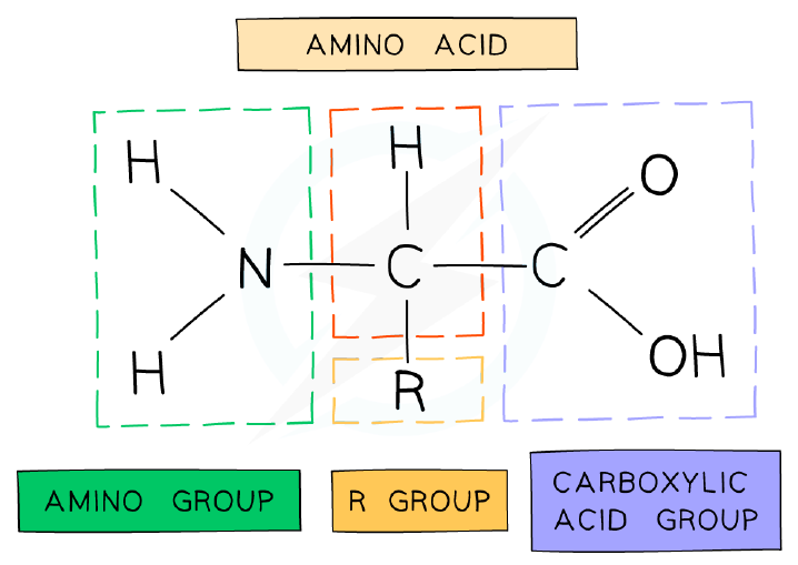
Amino Acid Structure
- Amino acids are the monomers of polypeptides; there are 20 amino acids common to all living organisms.
- Each amino acid has a central carbon atom (α carbon) bonded to an amine group (-NH₂), a carboxyl group (-COOH), a hydrogen atom, and an R group.
- The R group is the variable part that makes each amino acid different; it determines properties such as whether the amino acid is acidic or basic, or polar or non-polar.
- The R group can be as simple as a hydrogen atom (glycine) or as complex as an aromatic ring (phenylalanine).
- In a neutral environment, the carboxyl group donates a hydrogen ion (becoming negative) and the amine group accepts a hydrogen ion (becoming positive).
General structure of an amino acid
Peptide Bond Formation
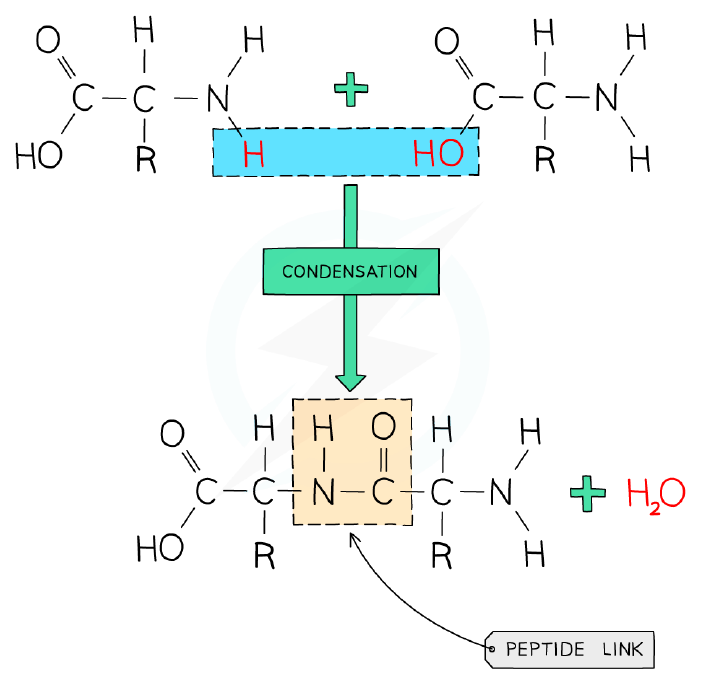
- A peptide bond forms between the carboxyl group of one amino acid and the amine group of another.
- During peptide bond formation, a hydroxyl group (-OH) is lost from the carboxyl group and a hydrogen atom is lost from the amine group.
- This is a condensation reaction, so water is released.
- Dipeptides form from two amino acids; polypeptides form from three or more amino acids.
- Hydrolysis breaks peptide bonds by adding water, splitting polypeptides back into amino acids.
- The R group is not involved in peptide bond formation.
Forming a polypeptide
Dietary Requirements for Amino Acids
- Humans can synthesise 11 non-essential amino acids from other amino acids.
- The remaining 9 essential amino acids must be obtained from the diet.
- A healthy, varied diet provides all essential amino acids; meat contains all nine, so vegetarian and vegan diets must be well balanced.
- Diets that restrict certain foods may require supplementation to ensure all essential amino acids are consumed.
The Variety of Proteins
- Protein diversity arises from the 20 amino acids, variation in polypeptide length (from a few to thousands), and differences in amino acid sequence.
- The DNA base sequence determines the number and order of amino acids in a polypeptide.
- For a polypeptide of 50 amino acids, there are 20⁵⁰ possible combinations (≈1.13 × 10⁶⁵); for an average protein of 300 amino acids, the number is effectively infinite.
- Polypeptides are assembled at ribosomes by adding amino acids one by one; the mRNA codon determines which amino acid is added.
- Proteins have many roles: enzymes (catalysis), blood clotting, structural fibres (collagen, keratin), transport (haemoglobin), cytoskeleton formation, cell adhesion, hormones, DNA compaction (histones), antibodies, membrane transport, and cell receptors.
Examples of Proteins
- Rubisco (ribulose bisphosphate carboxylase) is a globular enzyme with 16 polypeptide chains that fixes CO₂ in photosynthesis; it is the most abundant enzyme on Earth.
- Insulin is a short, globular hormone with 2 polypeptide chains, produced by β-cells in the pancreas; it binds to insulin receptors on liver, fat, and muscle cells to cause glucose absorption.
- Immunoglobulins (antibodies) are globular proteins with a Y shape and highly variable binding sites, allowing them to bind millions of different antigens; they are the most diverse group of proteins.
- Rhodopsin is a membrane protein in rod cells of the retina; it contains retinal (derived from vitamin A) and undergoes a conformational change when hit by a photon, sending a nerve impulse.
- Collagen is a fibrous protein of three polypeptide chains forming a triple helix; it is the most abundant protein in the human body (~25%) and provides tensile strength in skin, blood vessels, and connective tissue.
- Spider silk is a fibrous protein as strong as steel but lighter; it has rope-like and coiled parts, does not denature easily at extreme temperatures, and can be genetically engineered to be expressed in goats' milk.
Protein Structure: Effect of pH & Temperature
- Protein structure is sensitive to temperature and pH because these factors affect the ionic interactions, hydrogen bonds, and other intermolecular forces that maintain the 3D shape.
- Denaturation is the irreversible change of protein conformation; the bonds holding the shape are relatively weak and can be easily broken.
- Denaturation alters the protein's shape, which may affect its function, physical state, and usefulness.
- Each protein has an optimum pH at which its 3D structure is not denatured.
- Denaturation is almost always irreversible, but small denaturations and renaturations can occur in some proteins (e.g., haemoglobin) in response to small pH fluctuations.
- In egg white (albumin), heating causes hydrophobic amino acids to move to the edges, making the protein insoluble and forming a solid cooked layer.
- In the stomach, low pH (pH 2) denatures dietary proteins; the enzyme pepsin has an optimum pH of 2.
- Some extremophiles, such as Thermus aquaticus, have proteins stable at extreme temperatures (e.g., 80°C).
Levels of Protein Structure
- Primary structure is the sequence of amino acids in a polypeptide; the DNA determines this sequence, and the precise position of each amino acid determines the final 3D shape.
- Secondary structure arises from coiling or pleating of the amino acid chain, held together by hydrogen bonds between carbonyl (C=O) and amine (N-H) groups on parallel strands.
- Secondary structure includes α (α) helices and β (β)-pleated sheets.
- Tertiary structure is the complex 3D shape formed by folding of the secondary structure; it is determined by interactions between R groups.
- Interactions in tertiary structure include hydrogen bonds between polar R groups, hydrophobic interactions between non-polar R groups and water, disulfide bridges between cysteine R groups, and ionic bonds between charged R groups.
- Quaternary structure occurs when a protein consists of multiple polypeptide chains (subunits) functioning together; proteins with only one chain do not have quaternary structure.
- Quaternary proteins can be conjugated (containing non-protein prosthetic groups) or non-conjugated (no non-protein components).
- Haemoglobin is a conjugated protein with four polypeptide subunits, each containing a haem prosthetic group with an iron ion (Fe²⁺).
- Insulin (two subunits joined by disulfide bridges) and collagen (three subunits wound into a helix) are non-conjugated proteins with quaternary structure.
Globular & Fibrous Proteins
- Globular proteins are compact, roughly spherical, and soluble in water; they have hydrophobic R groups towards the centre and hydrophilic R groups on the outside.
- The solubility of globular proteins allows them to be transported and to function in metabolic reactions; their specific shapes enable roles such as enzymes and immunoglobulins.
- Some globular proteins are conjugated, e.g., haemoglobin contains the prosthetic group haem.
- Fibrous proteins are long strands of polypeptide chains with cross-linkages from hydrogen bonds; they have little or no tertiary structure.
- Fibrous proteins are insoluble in water due to a large number of hydrophobic R groups.
- Fibrous proteins have a highly repetitive amino acid sequence, creating strong, organised structures suitable for structural roles (e.g., keratin in hair and nails, collagen in connective tissue).
- Collagen is formed from three polypeptide chains held by hydrogen bonds into a triple helix, giving great tensile strength; covalent cross-links between R groups hold triple helices together to form fibrils.
- Collagen fibres are aligned with the forces they withstand; they are found in tendons, cartilage, ligaments, bones, teeth, skin, blood vessel walls, and the cornea.
Protein Imaging Technology
- Cryogenic electron microscopy (cryo-EM) rapidly freezes protein solutions and exposes them to electrons to produce images.
- Cryo-EM allows reconstruction of the 3D shape of proteins and visualisation of their interactions with other molecules.
- Unlike X-ray crystallography, cryo-EM does not require crystallisation, which can be time-consuming, may fail for some proteins, and removes contextual information by visualising proteins outside the cell.
スライド
練習問題
無料プレビュー — 57問中8問。すべて見るには登録を。
1.Which of the following is the monomer of a polypeptide?
Easy- AAmino acid
- BNucleotide
- CMonosaccharide
- DGlycerol
2.The following diagram of a dipeptide has four parts numbered 1 to 4. Part 1 is the group -NH2, part 2 is the group -COOH, part 3 is the bond joining the two amino acids, and part 4 is the variable side chain. Which row of the table correctly identifies each labelled part?
Easy- A1 = Amino group, 2 = Carboxyl group, 3 = Peptide bond, 4 = R-group
- B1 = Carboxyl group, 2 = Peptide bond, 3 = R-group, 4 = Amino group
- C1 = R-group, 2 = Amino group, 3 = Peptide bond, 4 = Carboxyl group
- D1 = Amino group, 2 = Peptide bond, 3 = R-group, 4 = Carboxyl group
3.Which of the following is not a function of proteins?
Easy- AActing as a store of chemical potential energy in cells
- BForming the cytoskeleton within a cell that facilitates chromosome movement
- CActing as chemical messengers that are secreted by glands and act on different parts of the body
- DSpeeding up chemical reactions within a cell
4.A simple tetrapeptide consists of four amino acids. How many different combinations of amino acids would be possible for this peptide?
Medium- A160 000
- B16 000
- C1 600 000
- D16 000 000
5.Which of the following are all examples of proteins?
Medium- ARhodopsin, immunoglobulins, rubisco
- BRubisco, collagen, amylopectin
- CAmylopectin, collagen, guanine
- DInsulin, amylose, spider silk
6.Which of the following is involved in the secondary structure of a protein? I. Double helix II. β-pleated sheets III. Hydrogen bonds IV. Hydrophobic interactions
Medium- AII and III
- BI and II
- CI, II and III
- DII, III and IV
7.Amino acids consist of oxygen, hydrogen, carbon and nitrogen atoms. Using bond-length data, the approximate length of one amino acid is 0.7 nm. What would be the approximate length of a dipeptide of this amino acid after a condensation reaction has occurred?
Medium- A1.2 nm
- B1.0 nm
- C1.4 nm
- D1.6 nm
8.Which of the following chemical groups does not bond directly with the central carbon of an amino acid?
Medium- A-OH
- B-NH2
- C-COOH
- D-H