Transport
Learn it by playing
Answer these questions to earn energy, then fish and explore. No account needed.
Lesson notes
Blood Vessels: Arteries
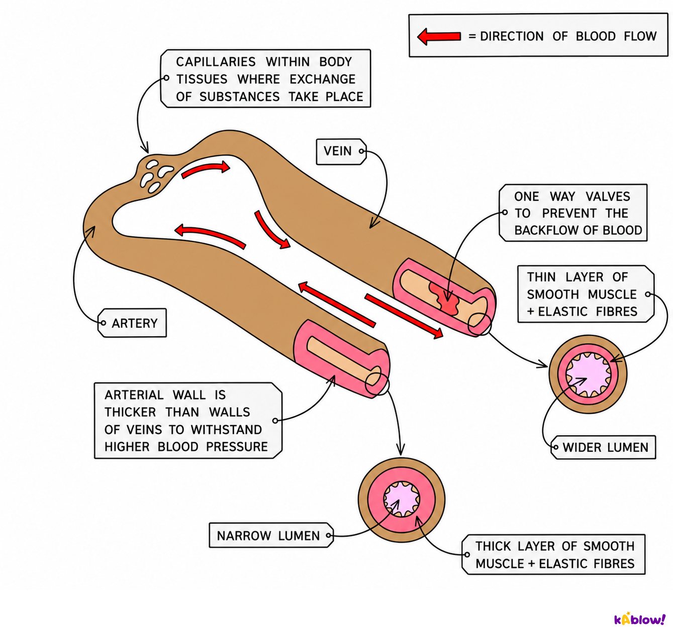
- Arteries transport blood away from the heart at high pressure.
- Artery walls have three layers: an inner endothelium (one cell thick, smooth, reduces friction), a middle layer of smooth muscle and elastic tissue, and an outer layer of collagen and elastic fibres.
- The thick muscle layer strengthens the artery to withstand high pressure and can contract or relax to regulate blood pressure by changing lumen diameter.
- Elastic tissue stretches and recoils to even out pressure fluctuations as the heart beats.
- Arteries have a narrow lumen which helps maintain high blood pressure.
- Systolic pressure is the peak pressure reached in arteries as blood is forced out of the ventricles; diastolic pressure is the lowest pressure as the heart relaxes.
- Vasoconstriction (decreased lumen diameter) increases blood pressure; vasodilation (increased lumen diameter) decreases it.
Comparing arteries and veins
Blood Vessels: Veins
- Veins transport blood to the heart at low pressure.
- Vein walls have a thin middle layer because they do not need to withstand high pressure.
- Veins have a wide lumen to maximise the volume of blood that can flow at any one time and reduce friction.
- Veins contain valves that prevent backflow of blood under low pressure.
- Movement of skeletal muscles pushes blood through veins; valves catch any backflow and the next muscle movement pushes it forward.
- Vein walls are flexible, allowing surrounding muscles and tissues to compress them and help move blood back to the heart.
The main blood vessels of the body
Blood Vessels: Capillaries
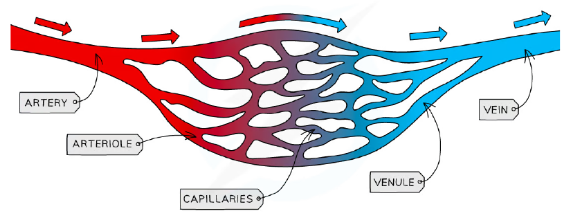
- Capillaries provide the exchange surface in tissues through networks called capillary beds.
- The wall is made of a single layer of endothelial cells, reducing the diffusion distance for oxygen and carbon dioxide.
- Some capillaries have gaps called fenestrations that allow blood plasma to leak out and form tissue fluid.
- Capillaries have a narrow lumen; red blood cells squeeze through in single file, slowing blood flow and providing more opportunity for diffusion.
- Capillaries branch between cells, increasing surface area for diffusion and reducing diffusion distance.
- The permeability of capillaries can vary depending on the requirements of a tissue.
The blood vessel network
Identifying Blood Vessels in Micrographs
- Arteries have thick walls with more collagen, elastic fibres and smooth muscle, and a narrow lumen relative to wall thickness.
- Veins have thin walls and a wide lumen relative to wall thickness.
- The wide lumen of veins reduces friction between blood and the endothelial layer.
- Blood flow is slower in veins, but the larger lumen means the volume of blood delivered per unit time is equal to that in arteries.
- In micrographs, distinguish arteries and veins by wall thickness and lumen diameter.
Measuring Pulse Rate
- Contraction of the ventricles forces blood through arteries, which expand; this can be felt as a pulse.
- The carotid artery (side of the neck, just below the jaw) or radial artery (wrist, below the base of the thumb) can be used to measure pulse rate.
- Place two fingers on the artery and gently compress it; count pulses for 60 seconds, or for 30 seconds and multiply by 2.
- Do not use your thumb because it has its own pulse and can give inaccurate results.
- Digital devices such as data loggers, smartwatches or fitness bands scan blood flow through the radial artery to measure pulse rate.
Monitoring heart rate
Coronary Heart Disease
- Occlusion is the narrowing of arteries due to a blockage.
- Atherosclerosis begins with damage to artery walls (e.g. from high blood pressure), leading to fatty deposits called atheromas under the endothelium.
- Atheromas narrow the lumen, reducing blood flow and increasing blood pressure, which causes further damage.
- Fibrous tissue repairs damage but is not elastic, reducing artery wall elasticity; the smooth lining breaks down forming plaques.
- Plaque rupture can lead to blood clotting; a clot within a vessel is a thrombus, and a circulating clot is an embolus.
- If an embolus blocks a coronary artery, part of the heart muscle dies, which may lead to a myocardial infarction (heart attack).
- Blockages in coronary arteries may be bypassed by heart bypass surgery, using blood vessels from the patient's leg to create an alternative route.
The coronary arteries
Evaluating Data on Coronary Heart Disease
- Epidemiological studies provide correlation data and do not provide a definite causal link between coronary heart disease and risk factors such as saturated fat intake.
- Clinical studies of individual patients are often small and may not be representative; they lack controlled experiments, so causal links cannot be made.
- Ethical considerations prevent controlled experiments that would require one group to consume a high-fat diet over a long period.
- When evaluating data, consider whether the sample group is representative of the population; larger sample sizes are more likely to be representative.
- Samples must not all come from the same demographic group, and samples must be human because animal trials do not perfectly represent human physiology.
- Statistical analysis (e.g. error bars, comparison of means) should be used to check that differences are statistically significant.
- Studies should be repeated or supported by many studies before conclusions are drawn; researchers should not be biased, and data collection methods must be accurate.
- Correlation is an association between variables; it does not necessarily imply causation.
- The correlation coefficient (r) indicates the strength of a relationship; perfect correlation is 1 or -1, and no correlation is 0.
The Transpiration Stream
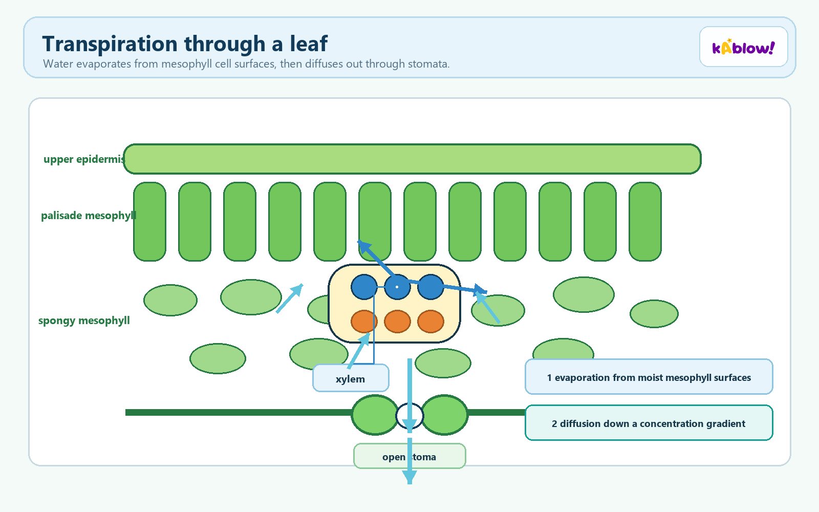
- Water evaporates from leaf cell surfaces during transpiration; more water is drawn from the nearest xylem vessels to replace it.
- Water molecules adhere to cell walls, enabling movement through cell walls; this movement due to adhesion in a narrow tube is capillary action.
- Loss of water from xylem generates tension (negative pressure) within the xylem.
- Tension creates a pulling force transmitted via cohesion between water molecules all the way down the stem to the roots.
- This is known as transpiration pull and allows water to move upwards against gravity; it is sometimes called the cohesion-tension theory.
- The continuous upwards flow of water in xylem vessels is the transpiration stream.
- Transpiration is important for cooling the plant via evaporative cooling, uptake of mineral ions, and providing turgor pressure for support of leaves and non-woody stems.
Transpiration through a leaf
Adaptations of Xylem Vessels
- Xylem vessels transport water and mineral ions from the roots.
- Xylem vessels are formed from long lines of cells connected end to end; mature vessels are non-living.
- Cell walls between connected cells degrade and cell contents are broken down, forming long, continuous, hollow tubes lacking cell content and end walls.
- This allows unimpeded flow through the xylem vessels.
- Walls are thickened with cellulose and strengthened with lignin, making them tough enough to withstand very low internal pressures (tension) without collapsing.
- Xylem vessel walls contain tiny pores called pits which allow water to enter and move sideways between vessels; if a vessel is damaged, water can flow into another vessel.
Vascular tissue in a plant
Drawing Root & Stem Structure
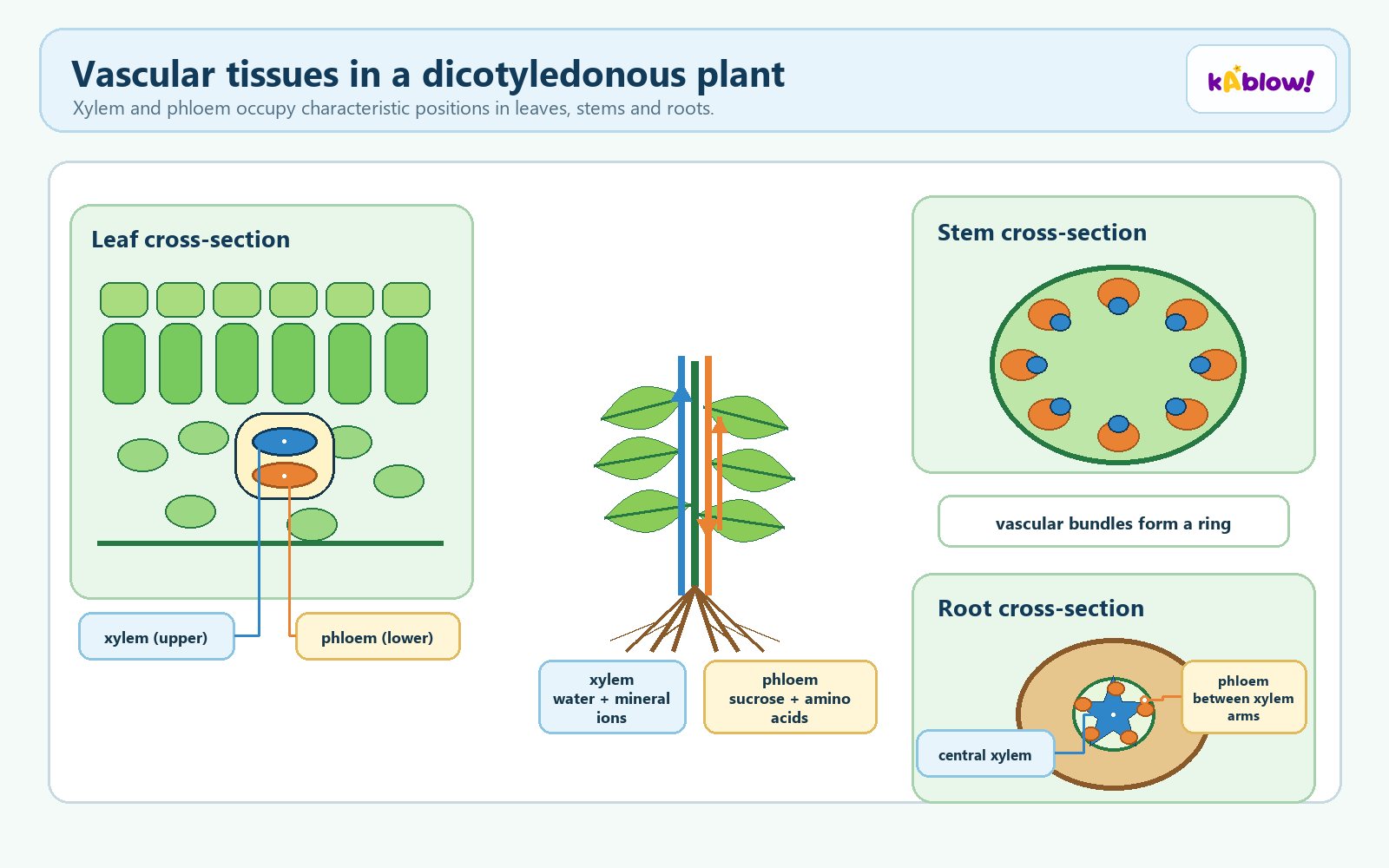
- In a dicotyledonous stem, tissues include the epidermis (outer layer, prevents water loss and protects from herbivores), parenchyma forming the cortex and pith (storage of starch and other substances), and vascular tissue arranged in a ring of vascular bundles.
- In the stem, xylem transports water and dissolved mineral ions from roots to leaves; phloem transports organic solutes from leaves to other parts.
- In a dicotyledonous root, xylem is centrally located in a cross-shaped structure; phloem bundles are arranged between the cross arms of xylem.
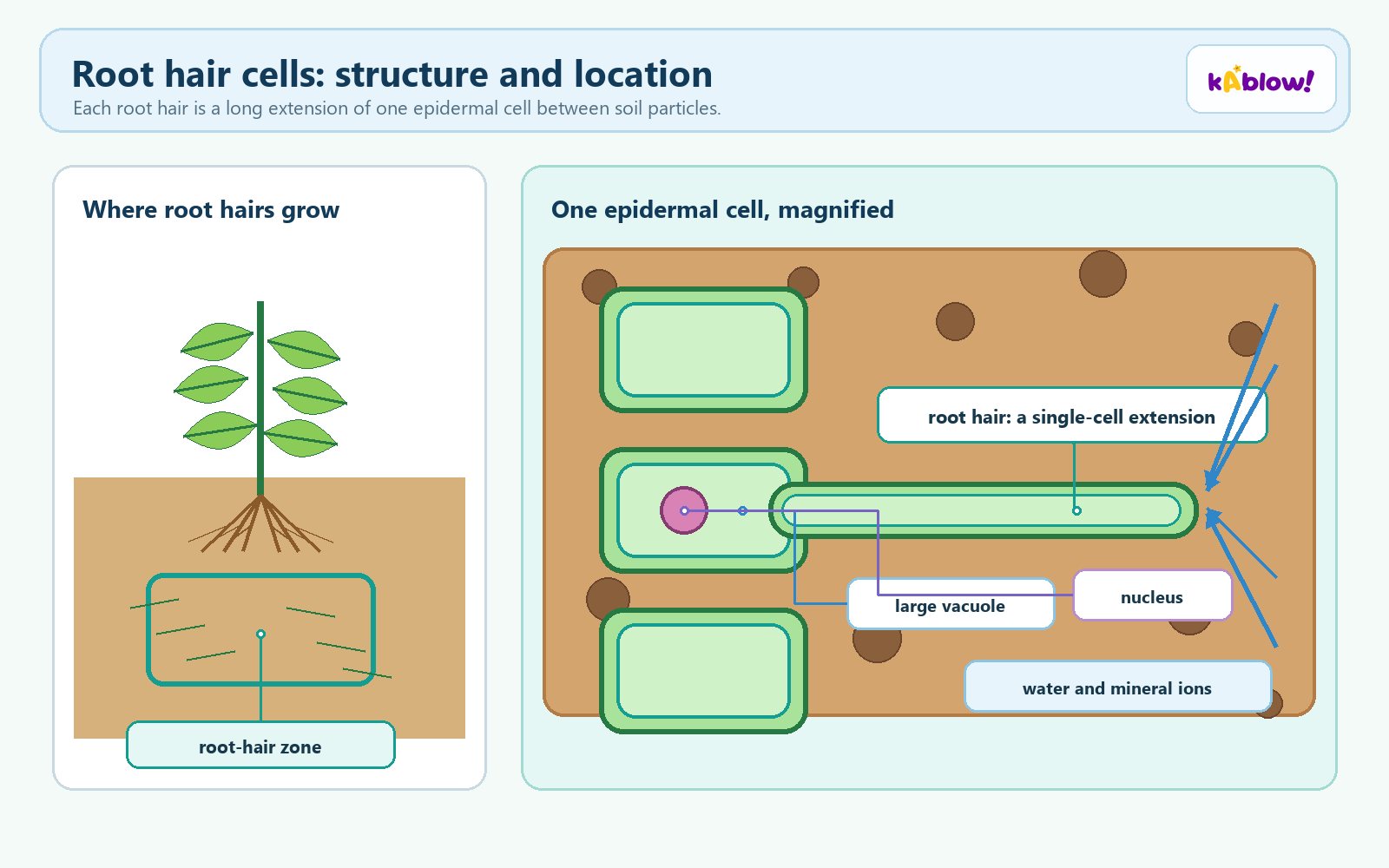
- The root cortex consists of parenchyma cells that store starch; the epidermis forms the outer layer and has root hairs for absorption of water and mineral ions.
- The endodermis forms the boundary between the vascular tissue and cortex in a root.
- When drawing plan diagrams: do not draw individual cells, use clear continuous lines, avoid shading, pay attention to proportions, label clearly, add a scale bar or estimated size, and annotate with functions.
- Draw plan diagrams large enough to fill at least half of the available space on a page.
Root hair cells: structure and location
Tissue Fluid
- Plasma is a straw-coloured liquid constituting about 55% of blood, largely water (95%), and is a good solvent for transport.
- As blood passes through capillaries, some plasma is forced out through gaps in capillary walls to surround body cells, forming tissue fluid.
- Tissue fluid contains fewer proteins and cells than plasma; proteins, red blood cells and platelets are too large to leave the capillaries.
- Exchange of substances between cells and blood occurs via tissue fluid; e.g. carbon dioxide from respiration dissolves into tissue fluid and moves back into the capillary.
- At the arterial end of a capillary, high blood pressure forces fluid out; this is pressure filtration.
- At the venous end, blood pressure is reduced and tissue fluid drains back into the capillaries; roughly 90% of fluid lost is reabsorbed.
- The remaining 10% of fluid is collected by lymph vessels and eventually returned to the circulatory system.
- After pressure filtration, proteins remain in the blood, creating a solute gradient that draws water back into the capillary by osmosis.
- At the arterial end, hydrostatic pressure (blood pressure) is greater than the solute gradient, so net movement is out; at the venous end, the solute gradient is greater, so fluid moves back in.
Lymphatic System
- Some tissue fluid enters lymph capillaries, which are separate from the circulatory system and form part of the lymphatic system.
- Lymph capillaries have closed ends and large pores that allow large molecules to pass through.
- Larger molecules that cannot pass through capillary walls enter the lymphatic system through small valves in the vessel walls.
- Lymph moves along larger vessels by compression caused by body movement; backflow is prevented by valves.
- Lymph nodes in the lymph ducts contain macrophages that engulf bacteria and other immune system cells.
- Lymph eventually re-enters the bloodstream through veins located close to the heart.
- Plasma proteins that escape from blood are returned via lymph capillaries; if not removed, they could increase solute concentration of tissue fluid and prevent water reabsorption.
- After digestion, lipids are transported from the intestines to the bloodstream by the lymph system, so lymph fluid contains lipids.
Circulatory Systems
- Both fish and mammals have a closed circulatory system, meaning blood is contained within blood vessels.
- Bony fish have single circulation: blood moves through the heart once per complete circuit; the heart has two chambers.
- In fish, blood enters the heart from the body, is pumped to the gills for oxygenation, then moves to the rest of the body.
- Mammals have double circulation: blood flows through the heart twice per complete circuit; the heart has four chambers.
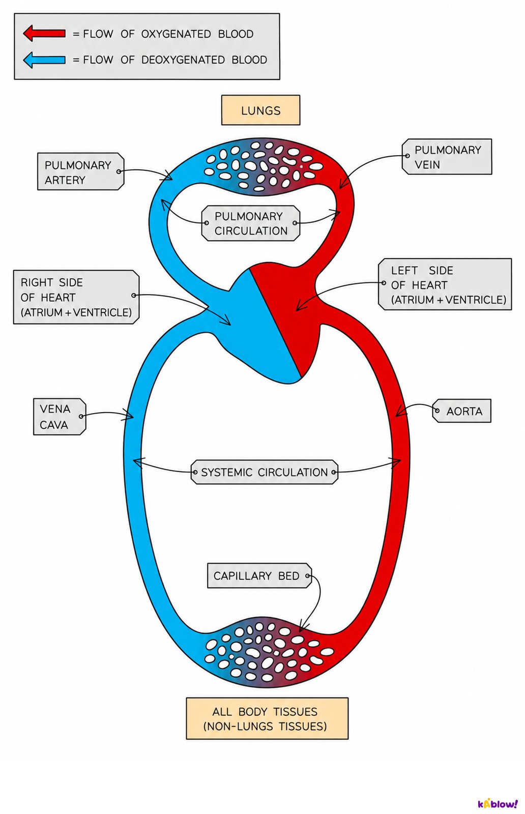
- The right side of the mammalian heart pumps deoxygenated blood to the lungs (pulmonary circulation); blood returns to the left side and is pumped around the body (systemic circulation).
- Advantages of double circulation: keeping oxygenated and deoxygenated blood separate, maintaining high pressure for transport to the body, and pumping blood at lower pressure in the lungs to avoid damaging delicate vessels.
Structure of the mammalian heart
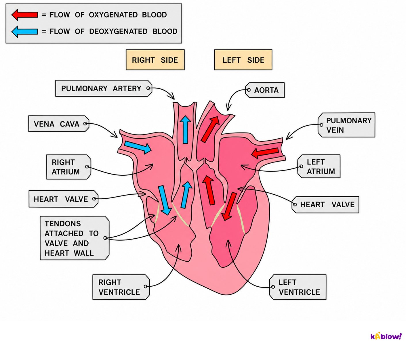
The Mammalian Heart
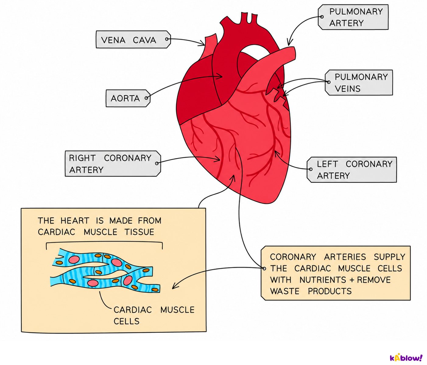
- The heart is made of cardiac muscle, which contracts repeatedly and continuously without fatigue.
- The heart has four chambers: two atria (top) and two ventricles (bottom).
- Atria receive blood from veins and have thin muscle walls, pumping blood a short distance into the ventricles.
- Ventricles receive blood from atria and have thick muscle walls to generate high pressure; the left ventricle has thicker muscle than the right because it pumps blood around the whole body.
- The septum is a wall of muscular tissue separating the left and right sides of the heart, ensuring blood does not mix.
- The pacemaker (sinoatrial node, SAN) in the right atrium initiates the heartbeat by sending a wave of excitation across the atria, causing atrial contraction; the impulse travels to the base of the ventricles and spreads upwards, causing ventricular contraction.
- Blood vessels bringing blood to the heart: vena cava and pulmonary vein; vessels taking blood away: pulmonary artery and aorta.
- Valves keep blood flowing forward: atrioventricular valves (tricuspid on right, bicuspid on left) prevent backflow from ventricles to atria; semilunar valves (pulmonary and aortic) prevent backflow from arteries to ventricles.
- Valves open when pressure behind them is greater than pressure in front, and close when pressure in front is greater than behind.
- The heart receives its own blood supply through coronary arteries, which supply oxygen and glucose for aerobic respiration.
Translocation in Plants
- The two main assimilates carried in phloem are sucrose and amino acids.
- A source is where assimilates are produced or loaded into the phloem (e.g. leaves); a sink is where they are unloaded and used or stored (e.g. root tubers, seeds).
- When sucrose reaches its sink, it is converted to starch.
- Starch is a compact, insoluble storage polysaccharide composed of glucose units.
- Once water in the translocation stream reaches a sink, changes in water potential cause water to move into the xylem and be transported back up the plant.
- Plasmodesmata in phloem cells tend to have a wider diameter than those in xylem cells, allowing larger molecules to pass between cells.
- Phloem sieve tubes minimise frictional resistance to flow by having few or no organelles and perforated sieve plates at their ends.
- Girdling (removing a complete ring of bark) removes phloem, preventing translocation of assimilates to the roots; the tree dies over time as roots are starved.
- Xylem and phloem differ: xylem transports water and mineral ions upwards, is composed of dead cells, has lignified walls and no end walls; phloem transports organic solutes bidirectionally, is composed of living cells, has sieve plates and companion cells.
Slides
Sign up free to view the lesson slides
Step through every slide for this topic — plus flashcards and revision notes — with a free account.
Practice questions
Free preview — 8 of 62 questions. Sign up to see them all.
1.Which blood vessel carries blood away from the heart?
Easy- AArtery
- BVein
- CCapillary
- DVenule
2.Which of the following is a characteristic of capillaries?
Easy- AWalls are one cell thick
- BWalls contain a thick layer of smooth muscle
- CThey have a wide lumen
- DThey contain valves to prevent backflow
3.What is the main function of valves in veins?
Medium- ATo prevent backflow of blood
- BTo increase blood pressure
- CTo slow down blood flow
- DTo filter blood
4.Which of the following is a consequence of atherosclerosis?
Medium- ANarrowing of arteries
- BWidening of arteries
- CDecreased blood pressure
- DIncreased elasticity of artery walls
5.What is the name of the vessel that carries oxygenated blood from the lungs to the heart?
Easy- APulmonary vein
- BPulmonary artery
- CAorta
- DVena cava
6.Which of the following are functions of the xylem? (select all that apply)
Medium- ATransport of water
- BTransport of mineral ions
- CTransport of sucrose
- DTransport of amino acids
- ESupport of the plant
7.Which of the following are adaptations of xylem vessels for transport? (select all that apply)
Medium- ALignin in walls
- BPits in walls
- CCompanion cells
- DNo end walls
- ESieve plates
8.The left ventricle has a thicker muscular wall than the right ventricle.
EasyTrue or false?
Unlock all 62 questions & more
Create a free account to see every question, the slides, flashcards and revision notes for this topic.