Hazards and uses of radioactive emissions and of background radiation
Aprenda jogando
Responda a estas perguntas para ganhar energia, depois pesque e explore. Sem precisar de conta.
Notas de aula
Background Radiation
- Background radiation is around us all of the time and comes from both natural and man-made sources.
- Natural sources include rocks and cosmic rays from space.
- Man-made sources include fallout from nuclear weapons testing and nuclear accidents.
- The level of background radiation and the radiation dose a person receives can be affected by their occupation and/or location.
- Some areas of the world have higher background radiation because they are closer to sources of radiation.
- All living things emit a small amount of radiation — the amount within a banana is tiny and not at all dangerous.
- Background count rate must be subtracted from measured count rates to give the corrected count rate.
Radiation Dose and Regulation
- The amount of radiation received by a person is called the dose, measured in sieverts (Sv).
- One sievert is a very big dose and would cause acute radiation poisoning.
- People normally receive about 3 mSv (0.003 Sv) in one year.
- The UK limit for nuclear industry employees is 20 mSv in one year.
- A dosemeter (radiation badge) is worn by radiographers and anyone working with radiation to track the dose they receive.
- Exposure is regulated because ionising radiation can damage cells and tissues.
Dangers of Radiation
- Ionising radiation can damage human cells and tissues, causing tissue damage and mutations.
- High-energy radiation such as gamma rays and X-rays can damage healthy tissue if not properly targeted.
- If atoms in a DNA strand are ionised, the DNA can be damaged; the cell may die or the DNA may be mutated when it reforms.
- If a mutated cell replicates, a tumour may form — this is cancer, a significant danger of radiation exposure.
- Acute radiation exposure can cause skin burns and reduce white blood cells, lowering the immune system and making a person more susceptible to infections.
Handling Radiation Safely
- Radioactive sources should be kept in a shielded container when not in use, such as a lead-lined box.
- Sources should be handled with gloves and tongs to increase the distance from the body.
- Protective clothing may be worn to prevent the body becoming contaminated.
- The time a radioactive source is used for should be limited.
- After using a source, hands should be washed, and the date and time of use recorded.
Contamination and Irradiation
- Contamination is the unwanted presence of materials containing radioactive atoms on other materials — the material becomes radioactive.
- Contamination often happens due to a radiation leak, and is almost always a mistake or accident.
- Irradiation is the process of exposing a material to α, β or gamma radiation; it does not make the material radioactive.
- Irradiation can kill living cells, so it is used to sterilise surgical equipment and food.
- Contamination is more dangerous than irradiation because it causes continuous exposure to radiation.
- Contamination is especially dangerous if a source gets inside the body, irradiating internal organs as it moves through.
- Lead-lined suits reduce irradiation by absorbing radiation, while airtight suits prevent contamination by stopping radioactive atoms entering the body.
Half-Life and Risk
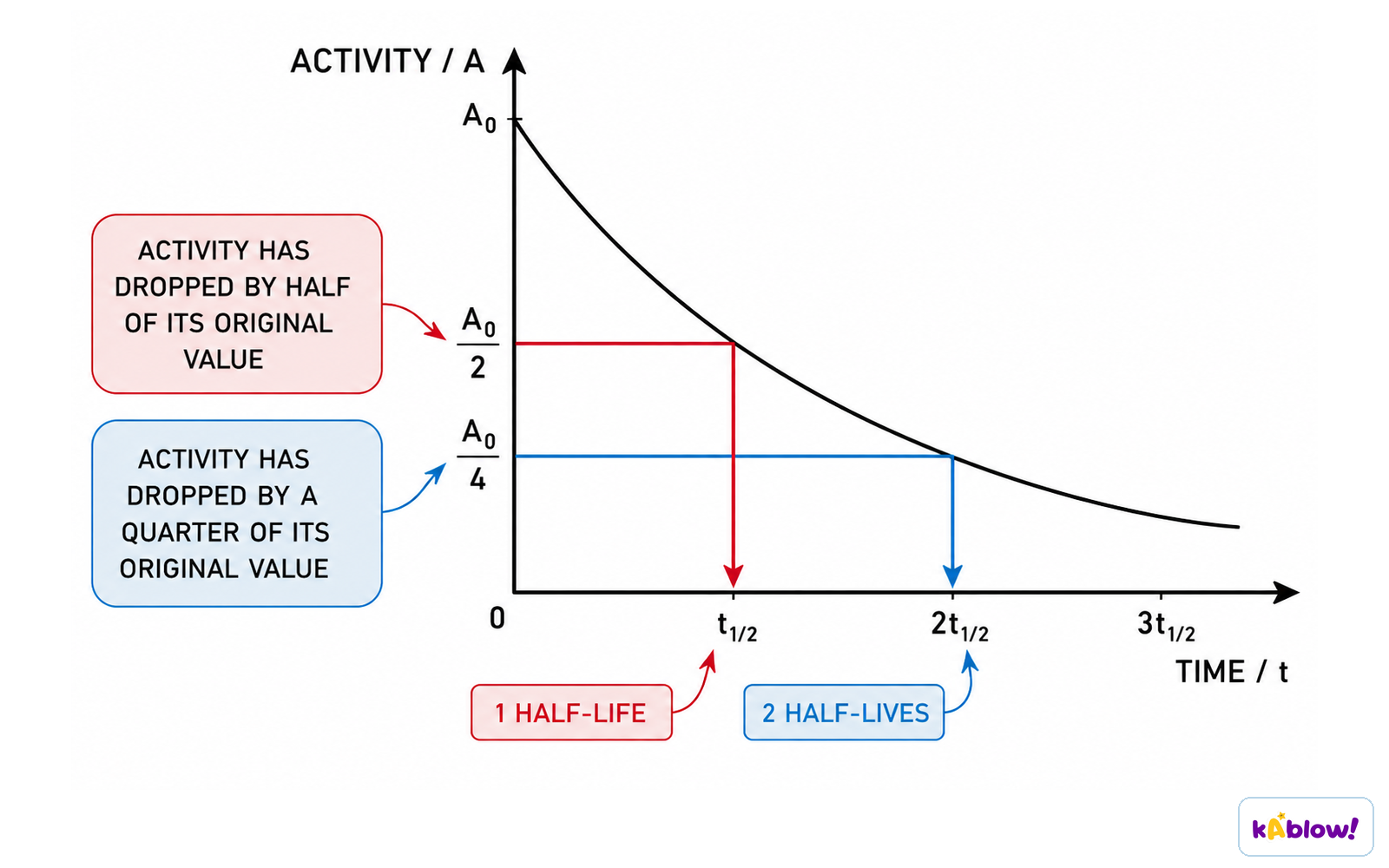
- The half-life is the time taken for the activity of a radioactive source to decrease to half of its original value.
- Different isotopes have very different half-lives: francium-218 is about 1 millisecond, polonium-210 about 140 days, and uranium-235 about 700 million years.
- A short half-life means nuclei decay quickly, emitting a lot of radiation in a short time — a greater risk of irradiation.
- If only a small amount is used, a short half-life is advantageous because the material quickly loses its radioactivity.
- A long half-life means a sample decays slowly but stays radioactive for a very long time — a greater risk of contamination.
- Radioactive waste with a long half-life is buried underground to prevent release into the environment.
A half-life graph
Medical Tracers
- A tracer is a radioactive isotope used to track the movement of substances, such as blood, around the body.
- Gamma emitters are usually used because gamma rays are highly penetrating and can pass out of the body to be detected externally, creating an internal image.
- Iodine-131 is an example of a radioactive tracer.
- Gamma rays are less ionising than some other radiation, so harm to the patient is minimised.
- The amount of isotope used is kept to a minimum to reduce exposure.
- Isotopes are chosen with short half-lives of around a few hours — long enough for the procedure but not so long as to cause long-term harm.
- Because of short half-lives, isotopes must be produced nearby or they would decay too much before reaching the hospital.
PET Scanning
- In PET scanning, positrons are emitted by the decay of the tracer.
- The positrons travel a small distance and annihilate when they interact with electrons in the tissue.
- This annihilation produces a pair of gamma rays (gamma photons) which can be detected outside the body.
- PET scanners are used to diagnose cancer and determine the location of a tumour.
Radiotherapy
- Radiotherapy is the treatment of cancer using radiation (chemotherapy uses chemicals).
- Radiation can kill living cells, and some cells — such as bacteria and cancer cells — are more susceptible than others.
- During external radiotherapy, beams of gamma rays are directed at the tumour; gamma is used because it can penetrate the body to reach the tumour.
- The beams are moved around to minimise harm to healthy tissue while still targeting the tumour.
- Surrounding healthy tissue is often shielded to avoid damage.
- During internal radiotherapy, small pellets of radioactive material are inserted into the tumour, exposing it directly to radiation.
Other Uses of Radiation
- α particles are used in smoke detectors: they ionise the air to create a current, and when smoke blocks the α emitter the current stops and the alarm triggers.
- β particles are used to measure the thickness of thin materials such as paper, cardboard or aluminium foil.
- β is used because it is partially absorbed — if α were used it would all be absorbed, and if gamma were used almost all would pass through with no detectable difference.
- As material gets thicker, more β particles are absorbed and fewer reach the detector; if it gets thinner, the opposite happens, allowing thickness to be kept constant.
- Gamma radiation is used to sterilise medical equipment because it is the most penetrating, irradiating all sides of instruments — even through packaging.
- Food can be irradiated to kill microorganisms, making it last longer and reducing food-borne infections.
- Radiation is also used to determine the age of ancient artefacts.
Evaluating Risk and Peer Review
- The hazards of radioactive material differ according to the half-life involved.
- The use of radiation in medicine carries risk, but the benefits can outweigh the risks — the risks from radiation are smaller than the risks of leaving a condition untreated.
- When evaluating risk, compare the potential dangers with the benefits, using given data.
- Some people worry about the safety and nutritional value of irradiated food.
- Scientific communities must be independent of government influence so their conclusions are trusted.
- Restaurants must clearly mark meals containing irradiated ingredients so people can make informed choices.
- Peer review is important when publishing findings on radiation risk.
Slides
Sign up free to view the lesson slides
Step through every slide for this topic — plus flashcards and revision notes — with a free account.
Questões de prática
Prévia grátis — 8 de 63 perguntas. Cadastre-se para ver todas.
1.What is meant by the term 'background radiation'?
Easy- ARadiation that is present in the environment all of the time
- BRadiation emitted only by nuclear power stations
- CRadiation given to a patient during a medical scan
- DRadiation that is deliberately released during a nuclear accident
2.Which of the following are sources of background radiation? (select all that apply)
Medium- ARocks
- BCosmic rays from space
- CFallout from nuclear weapons testing
- DA medical tracer injected into a patient
- EA smoke detector in a home
3.All background radiation is man-made.
EasyTrue or false?
4.Which of the following is a man-made source of background radiation?
Easy- ARadon gas from rocks
- BCosmic rays from space
- CFallout from nuclear weapons testing
- DFood and drink
5.Which statement correctly describes contamination?
Medium- AThe unwanted presence of materials containing radioactive atoms on other materials
- BThe process of exposing a material to α, β or gamma radiation
- CThe time taken for the activity of a radioactive source to halve
- DThe amount of radiation received by a person
6.Irradiating a material makes that material radioactive.
EasyTrue or false?
7.Which of the following is the correct definition of irradiation?
Medium- AThe process of exposing a material to α, β or gamma radiation
- BThe unwanted presence of radioactive atoms on a material
- CThe time taken for the activity of a source to halve
- DThe amount of radiation a person receives
8.Match each term to its correct definition.
Medium- Contamination
- Irradiation
- Half-life
- The process of exposing a material to α, β or gamma radiation
- The time taken for the activity of a radioactive source to halve
- The unwanted presence of materials containing radioactive atoms on other materials
Unlock all 63 questions, flashcards & more
Crie uma conta grátis para ver todas as perguntas, os slides, flashcards e resumos de revisão deste tópico.