The heart and blood circulation
Aprenda jogando
Responda a estas perguntas para ganhar energia, depois pesque e explore. Sem precisar de conta.
Notas de aula
Big idea: a transport system for the whole body
- Big idea (key concept): Systems. The heart, the blood vessels and the blood make up the circulatory system. Each part has its own job, and together they move materials around the body.
- Related concept: Transport. A cell in your toe cannot get oxygen straight from the air. The circulatory system carries substances to every cell and carries waste away.
- Global context: Identities and relationships. Your heart beats about 100 000 times every day, and the way you live, including how much you exercise, what you eat and whether you smoke, changes how well it works.
- Cells need oxygen and glucose for respiration, and they make waste such as carbon dioxide and urea. In a large animal these substances cannot simply diffuse from the surface to the middle of the body, because diffusion is far too slow over long distances.
- So humans have a transport system: the heart is the pump, the blood vessels are the tubes and the blood is the liquid that carries things.
- Humans have a closed circulatory system. Blood always stays inside blood vessels. Substances only pass in and out through the very thin walls of the smallest vessels, the capillaries.
The heart: chambers, valves and vessels
- The heart is a hollow organ made of cardiac muscle. It contracts and relaxes in a steady rhythm, about 70 times a minute in a person at rest.
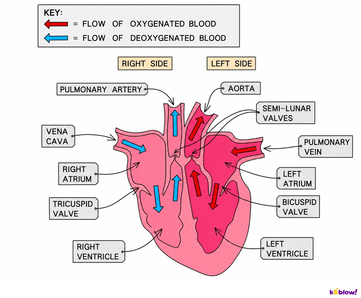
- It has four chambers. The two upper chambers are the atria (one is an atrium) and they receive blood coming into the heart. The two lower chambers are the ventricles and they pump blood out of the heart.
- A wall of muscle called the septum separates the right side from the left side, so blood with a lot of oxygen never mixes with blood that has little.
- The right side gets deoxygenated blood from the body through the vena cava into the right atrium. The right ventricle pumps it through the pulmonary artery to the lungs.
- The left side gets oxygenated blood from the lungs through the pulmonary vein into the left atrium. The left ventricle pumps it through the aorta to the rest of the body.
- The wall of the left ventricle is much thicker than the right. It has to push blood all the way round the body, while the right ventricle only pushes blood to the lungs, which are close by.
- Valves open in one direction only. The tricuspid valve (right) and bicuspid valve (left) lie between atria and ventricles, and semi-lunar valves guard the exits into the arteries. They stop blood flowing backwards, and the 'lub-dub' sound of a heartbeat is the sound of valves closing.
Inside the heart: chambers, valves and the direction of blood flow
Double circulation: two circuits, one heart
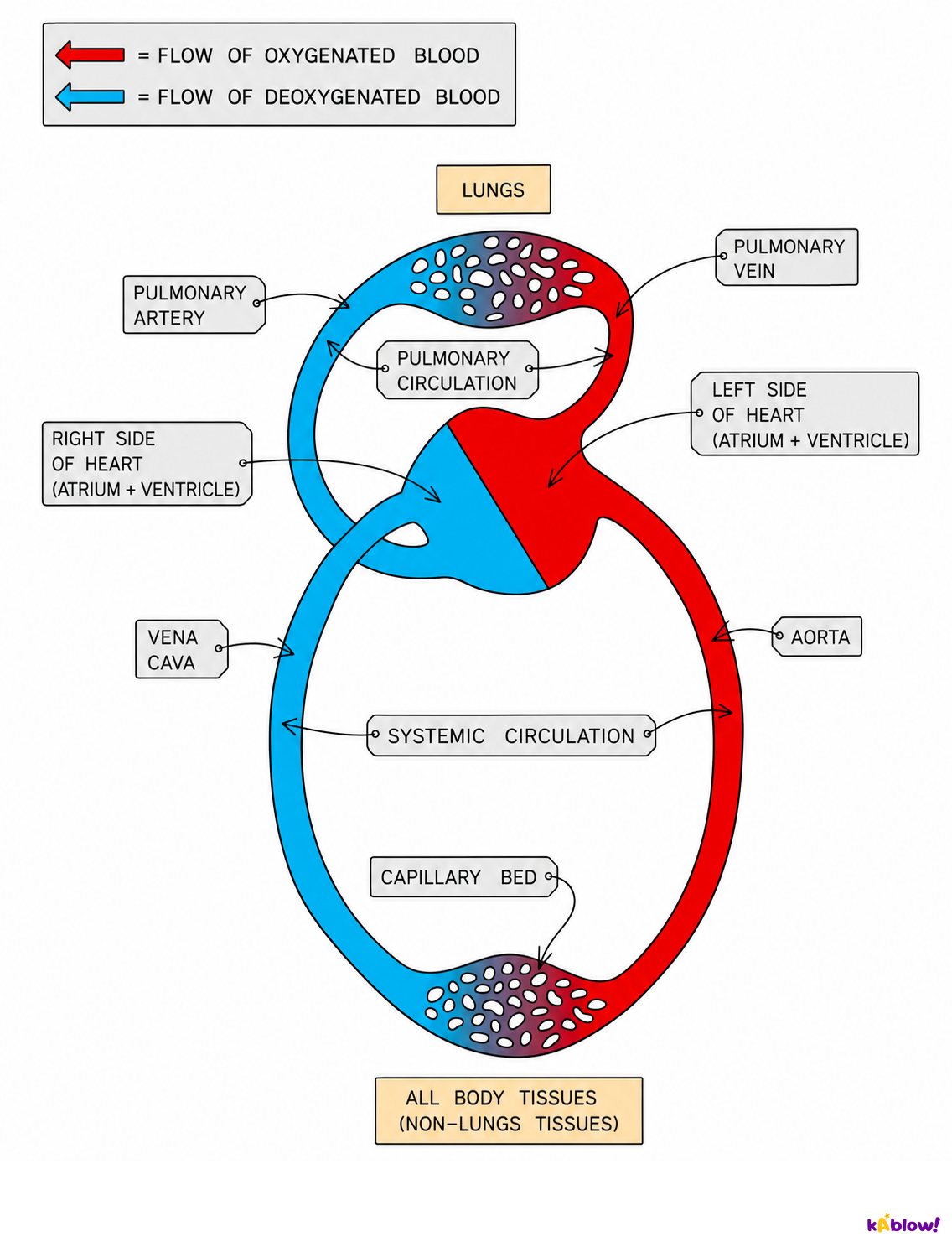
- Blood travels round two circuits. In the pulmonary circulation it goes from the heart to the lungs and back. In the systemic circulation it goes from the heart to the rest of the body and back.
- In the lungs, blood collects oxygen and loses carbon dioxide. It returns to the left atrium through the pulmonary vein.
- From the left ventricle, blood leaves through the aorta, travels through arteries to the organs, gives up oxygen in capillaries, and returns through veins and the vena cava to the right atrium.
- Blood passes through the heart twice on every complete trip round the body. That is why it is called a double circulation.
- Why is it useful? When blood is squeezed through the tiny capillaries in the lungs, its pressure drops. Returning to the heart lets the left ventricle give it a second push, so it reaches the body at high pressure.
- Keeping oxygenated and deoxygenated blood apart also means the body tissues receive blood carrying as much oxygen as possible. A fish has a single circulation, so blood reaches its body at a lower pressure.
The double circulation: lungs circuit and body circuit
Blood vessels: arteries, veins and capillaries
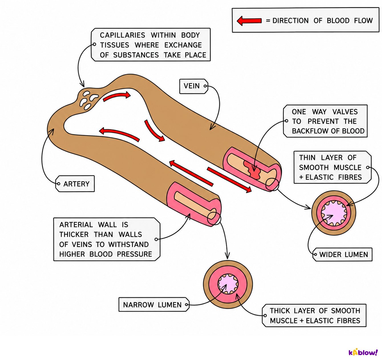
- Blood travels through three kinds of vessel. Arteries carry blood away from the heart, veins carry blood towards the heart and capillaries link the two.
- Arteries have thick walls with muscle and elastic fibres, and a narrow lumen (the space inside). They carry blood at high pressure. Their elastic walls stretch with each heartbeat and spring back, which you feel as the pulse.
- Veins have thinner walls and a wider lumen. Blood in them is at low pressure, so veins have valves to stop it flowing backwards. Muscles around the veins squeeze blood along when you move.
- Capillaries are tiny vessels with walls only one cell thick. Oxygen and glucose diffuse out through the wall to the cells, and carbon dioxide and waste diffuse in. Every living cell is close to a capillary.
- Blood leaves an artery through smaller arterioles, flows through capillaries, then joins venules that merge into veins.
- Do not decide by colour or by oxygen. Arteries usually carry oxygenated blood, but the pulmonary artery carries deoxygenated blood and the pulmonary vein carries oxygenated blood. Always decide by direction: away from the heart or towards it.
An artery and a vein compared
Blood: plasma and cells
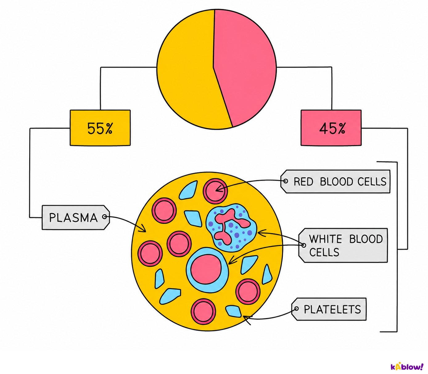
- About 55% of blood is plasma and about 45% is cells.
- Plasma is a pale yellow liquid, mostly water. It carries dissolved glucose, amino acids, carbon dioxide, urea, hormones and heat around the body.
- Red blood cells carry oxygen. They contain haemoglobin, a red protein that joins with oxygen in the lungs and releases it in the tissues. They have no nucleus, which leaves more room for haemoglobin, and their dent-in disc shape gives a large surface area for taking in oxygen.
- White blood cells defend the body against pathogens. Some, called phagocytes, engulf and digest pathogens. Others make antibodies.
- Platelets are tiny fragments of cells. They help the blood clot at a wound, which seals it and stops pathogens getting in.
What blood is made of
Think like a scientist: pulse, exercise and a healthy heart
- Each time the left ventricle contracts, a surge of blood stretches the arteries. You can feel this as a pulse at your wrist or neck. Your pulse rate is the same as your heart rate, measured in beats per minute (bpm). Most young people have a resting heart rate of about 60 to 100 bpm.
- When you exercise, your muscles respire faster. They need more oxygen and glucose and must get rid of more carbon dioxide, so your heart rate and breathing rate go up. The faster heart sends more blood to the muscles each minute. Fitter people usually have a lower resting pulse and recover faster after exercise.
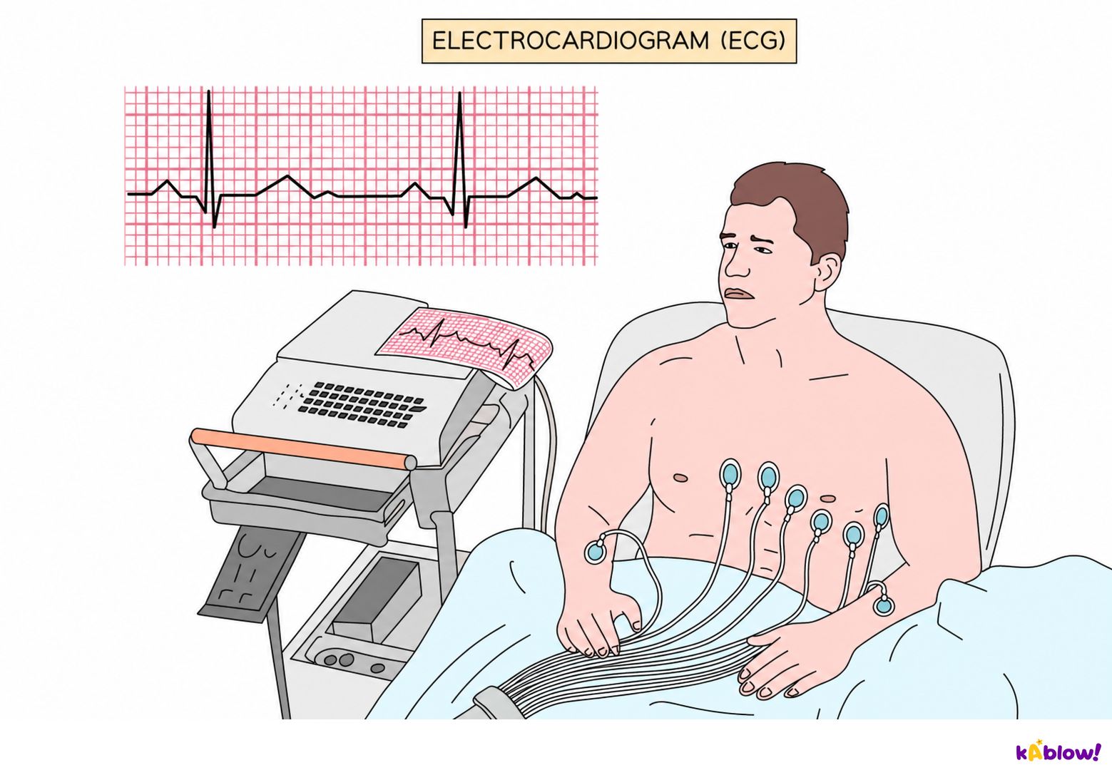
- An ECG (electrocardiogram) records the heart's electrical activity. Doctors use it to check heart rate and rhythm.
- The heart muscle has its own blood supply, the coronary arteries. In coronary heart disease fatty deposits narrow these arteries, so less oxygen reaches the heart muscle. A blockage can cause a heart attack. Smoking, a diet high in saturated fat and too little exercise raise the risk.
- The independent variable is the one you change on purpose (for example, how long a person jogs). The dependent variable is the one you measure (the pulse rate). A fair test keeps every other variable the same, and repeating measurements lets you take a mean.
- Inquiry task: plan an investigation into how the time spent exercising changes pulse rate. Say what you will change, what you will measure and how, name three variables you will control, and explain how you will make your results reliable. Then suggest one way to improve your method.
Recording the heart with an ECG
Slides
Sign up free to view the lesson slides
Step through every slide for this topic — plus flashcards and revision notes — with a free account.
Questões de prática
Prévia grátis — 8 de 50 perguntas. Cadastre-se para ver todas.
1.How many chambers does the human heart have?
Easy- ATwo
- BThree
- CFour
- DSix
2.Which chambers of the heart receive blood coming into the heart?
Easy- AThe atria
- BThe ventricles
- CThe valves
- DThe arteries
3.Which chambers of the heart pump blood out of the heart?
Easy- AThe atria
- BThe capillaries
- CThe veins
- DThe ventricles
4.Which blood vessel carries blood from the heart to the lungs?
Easy- AThe pulmonary vein
- BThe pulmonary artery
- CThe aorta
- DThe vena cava
5.Which blood vessel carries oxygenated blood from the lungs back to the heart?
Easy- AThe pulmonary artery
- BThe pulmonary vein
- CThe vena cava
- DThe aorta
6.What is the name of the large artery that carries oxygenated blood from the heart to the body?
Easy- AThe vena cava
- BThe pulmonary vein
- CThe capillary
- DThe aorta
7.What is the job of the valves in the heart?
Easy- ATo stop blood flowing backwards
- BTo pump blood out of the heart
- CTo add oxygen to the blood
- DTo make red blood cells
8.Which type of blood vessel carries blood away from the heart?
Easy- AVeins
- BCapillaries
- CArteries
- DValves
Unlock all 50 questions, flashcards & more
Crie uma conta grátis para ver todas as perguntas, os slides, flashcards e resumos de revisão deste tópico.