Circulatory Systems, Heart & Blood Vessels
Aprenda jogando
Responda a estas perguntas para ganhar energia, depois pesque e explore. Sem precisar de conta.
Notas de aula
Circulatory System Overview
- The circulatory system is an organ system that transports blood around the body.
- Components include blood vessels, a pump (heart), and valves for one-way flow.
- Fish have a two-chambered heart and single circulation (blood passes through heart once per circuit).
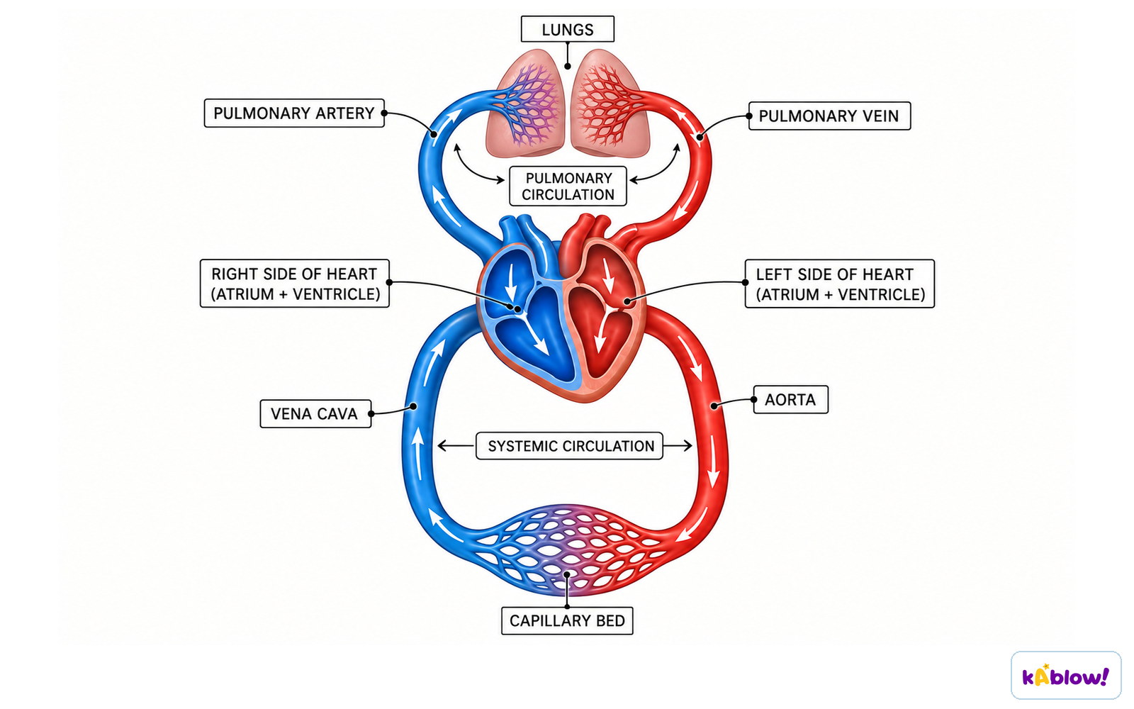
- Mammals have a four-chambered heart and double circulation (blood passes through heart twice per circuit).
- Double circulation: right side pumps deoxygenated blood to lungs (pulmonary circulation); left side pumps oxygenated blood to body (systemic circulation).
- Advantage: blood pressure lost in lung capillaries is restored by the heart before blood travels to the body, ensuring faster delivery of oxygen and glucose.
The double circulatory system
The Mammalian Heart
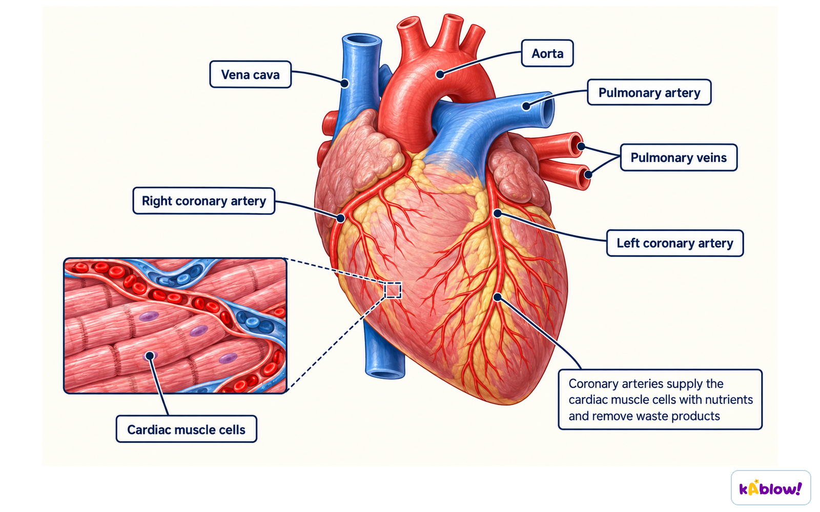
- The heart is a muscular pump; its own blood supply comes from the coronary arteries.
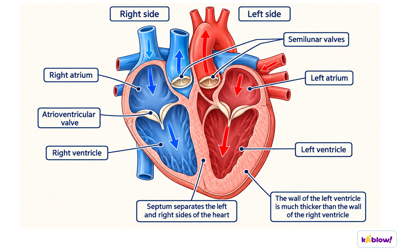
- The septum separates the right and left sides, preventing mixing of oxygenated and deoxygenated blood.
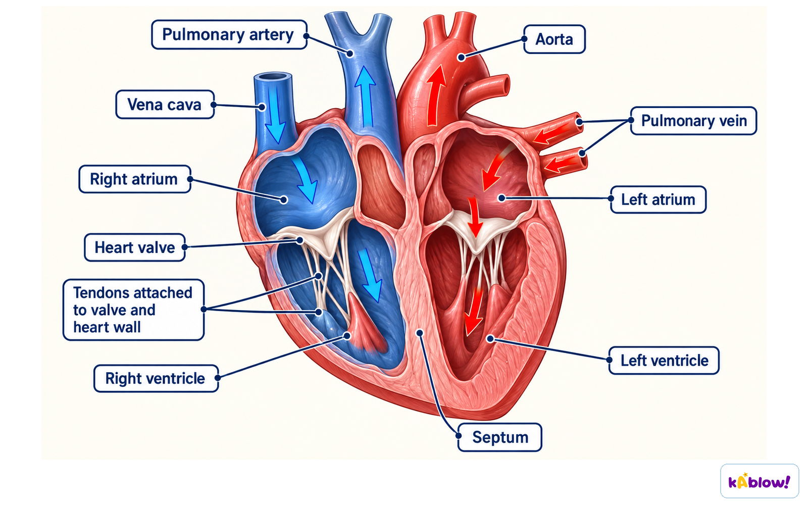
- Right side: receives deoxygenated blood from body via vena cava; pumps it to lungs via pulmonary artery.
- Left side: receives oxygenated blood from lungs via pulmonary vein; pumps it to body via aorta.
- Atria have thinner walls; ventricles have thicker walls (left ventricle thickest as it pumps to whole body).
- Valves: atrioventricular valves (tricuspid on right, bicuspid on left) prevent backflow into atria; semilunar valves in pulmonary artery and aorta prevent backflow into ventricles.
Structure of the mammalian heart
Heart Function & Cardiac Cycle
- Deoxygenated blood enters right atrium → passes through tricuspid valve into right ventricle → ventricle contracts → blood pushed through semilunar valve into pulmonary artery → to lungs.
- Oxygenated blood returns from lungs via pulmonary vein → enters left atrium → passes through bicuspid valve into left ventricle → ventricle contracts → blood pushed through semilunar valve into aorta → to body.
- Atrioventricular valves open when atria contract; close when ventricles contract to prevent backflow.
- Semilunar valves open when ventricles contract; close to prevent blood flowing back into heart.
Heart valves and septum
Monitoring Heart Activity & Exercise
- Heart activity monitored by ECG, pulse rate (bpm), or listening to valve sounds with a stethoscope.
- During exercise, heart rate increases to deliver more oxygen and glucose to muscles and remove waste faster.
- After exercise, heart rate remains elevated to remove excess waste and repay oxygen debt (break down lactic acid).
- Pulse rate gradually returns to resting level over several minutes.
Coronary Heart Disease (CHD)
- Coronary arteries supply oxygen and nutrients to heart muscle.
- CHD occurs when these arteries become narrowed or blocked by fatty plaques (mainly cholesterol).
- Partial blockage causes angina (chest pain); complete blockage leads to heart attack (muscle cells die).
- Risk factors: smoking, high-fat diet, lack of exercise, obesity, high blood pressure.
- Prevention: quit smoking, eat less animal fat, exercise regularly.
The coronary arteries
Blood Vessels: Arteries, Veins, Capillaries
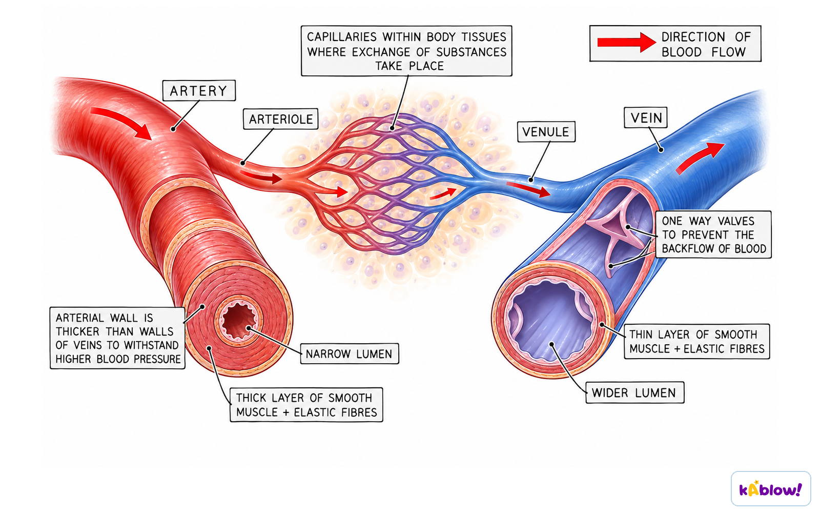
- Arteries carry blood away from heart at high pressure; thick muscular walls with elastic fibres; narrow lumen; mostly oxygenated (except pulmonary artery).
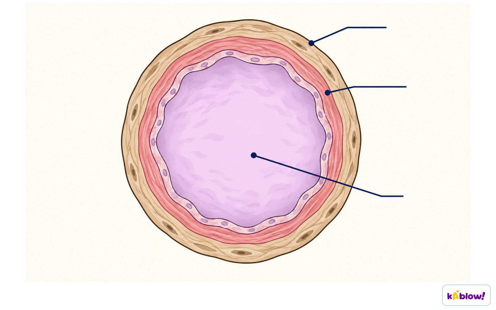
- Veins carry blood towards heart at low pressure; thin walls; wide lumen; contain valves to prevent backflow; mostly deoxygenated (except pulmonary vein).
- Capillaries are one cell thick with 'leaky' walls for diffusion; very narrow lumen; connect arterioles to venules.
- Arteries divide into arterioles then capillaries; capillaries merge into venules then veins.
Adaptations of blood vessels
Circulation Around the Body
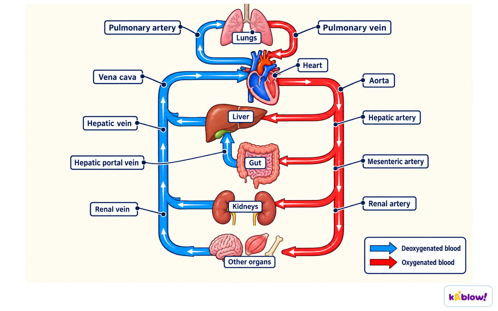
- Key blood vessels: aorta (to body), vena cava (from body), pulmonary artery (to lungs), pulmonary vein (from lungs).
- Renal artery carries blood to kidney; renal vein carries blood from kidney.
- Hepatic artery brings oxygenated blood to liver; hepatic portal vein brings deoxygenated blood from gut to liver; hepatic vein carries blood away from liver to heart.
Circulation around the body
Components of Blood
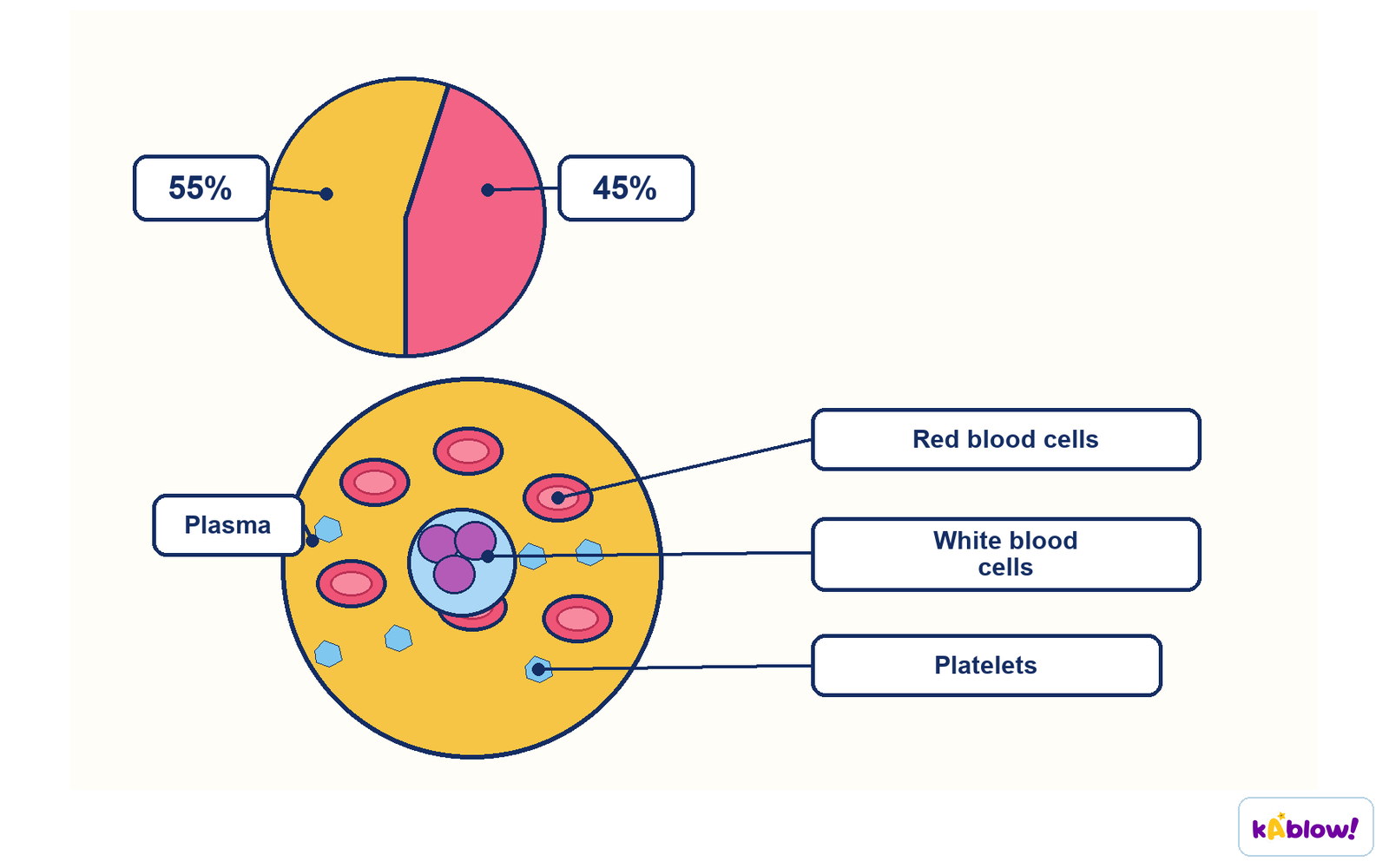
- Blood consists of plasma, red blood cells, white blood cells, and platelets.
- Plasma transports carbon dioxide, digested nutrients, urea, hormones, and heat.
- Red blood cells contain haemoglobin and transport oxygen as oxyhaemoglobin; no nucleus; biconcave disc shape.
- White blood cells defend against infection: phagocytes engulf pathogens (phagocytosis); lymphocytes produce antibodies and antitoxins.
- Platelets are cell fragments involved in blood clotting.
Composition of blood
Blood Clotting
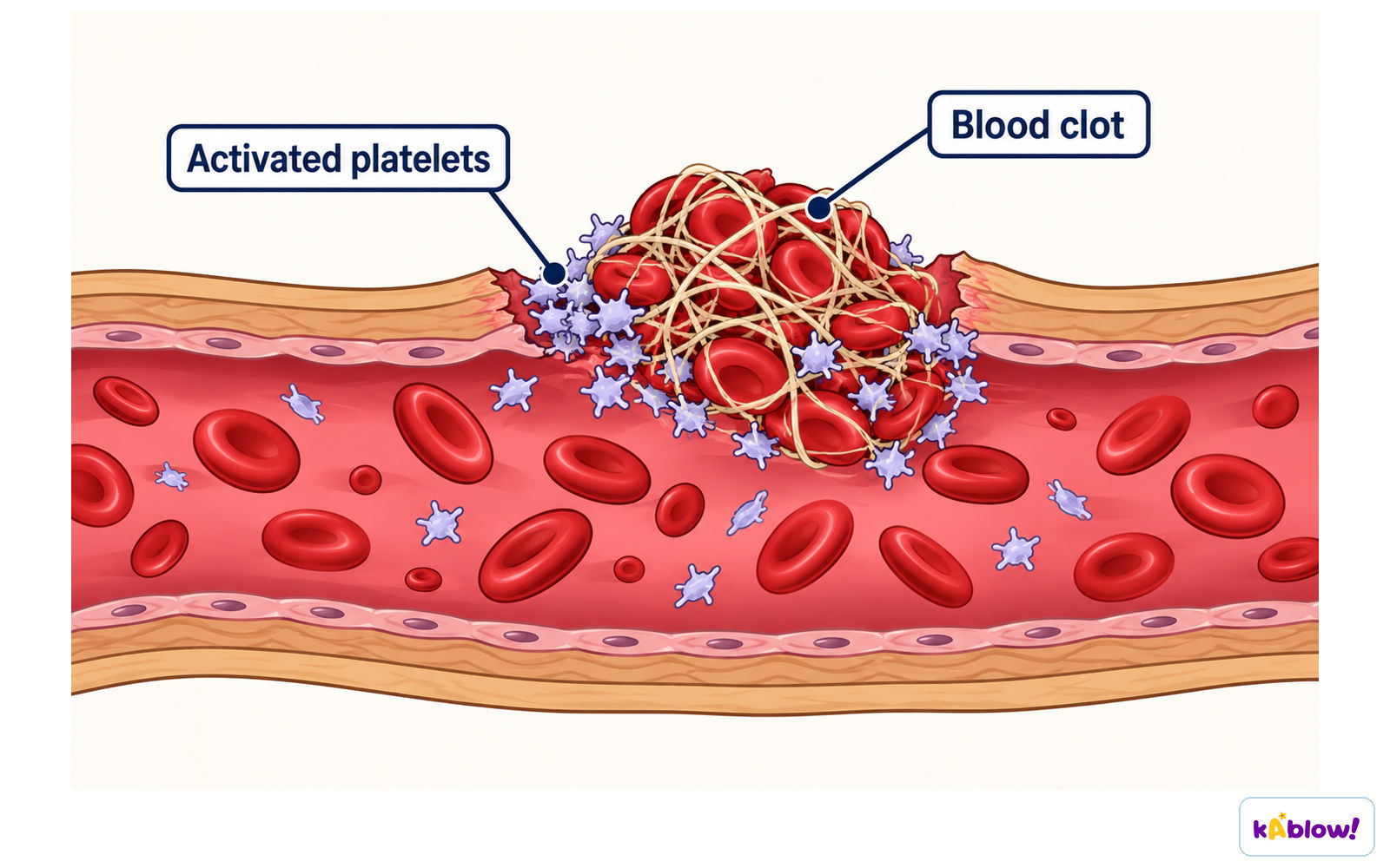
- When skin is broken, platelets release chemicals that convert soluble fibrinogen into insoluble fibrin.
- Fibrin forms a mesh that traps red blood cells, creating a clot.
- The clot dries to form a scab, preventing blood loss and entry of pathogens.
Blood clotting
Slides
Sign up free to view the lesson slides
Step through every slide for this topic — plus flashcards and revision notes — with a free account.
Questões de prática
Prévia grátis — 8 de 51 perguntas. Cadastre-se para ver todas.
1.Which sequence lists the heart chambers in order from thinnest wall to thickest wall?
Medium- Aleft ventricle → right ventricle → left atrium
- Bleft atrium → left ventricle → right ventricle
- Cright ventricle → left atrium → left ventricle
- Dleft atrium → right ventricle → left ventricle
2.Which of the following statements would be incorrect for most of the veins in the human body?
Easy- AThey carry blood at low pressure.
- BThey have valves.
- CThey have a pulse.
- DThey return blood to the heart.
3.The blood contains lymphocytes. What is their purpose?
Easy- ATransport of hormones
- BPhagocytosis
- CAntibody production
- DBlood clotting
4.Which of the following would be found dissolved in the blood plasma of a human?
Medium- AGlucose, urea and hormones
- BOxygen, urea and starch
- CCarbon dioxide, hormones and haemoglobin
- DCarbon dioxide, oxygen and urea
5.In which list of the following do all three blood vessels carry deoxygenated blood?
Medium- Aaorta, pulmonary vein, renal artery
- Bvena cava, pulmonary vein, renal vein
- Cvena cava, pulmonary artery, renal vein
- Daorta, pulmonary artery, renal artery
6.What happens to the heart valves when the ventricles contract?
Medium- Aatrioventricular valves close, semilunar valves close
- Batrioventricular valves close, semilunar valves open
- Catrioventricular valves open, semilunar valves close
- Datrioventricular valves open, semilunar valves open
7.Which part of the blood contains haemoglobin?
Easy- Aplasma
- Bplatelets
- Cred blood cells
- Dwhite blood cells
8.The diagram shows a cross-section through a human blood vessel. Which type of blood vessel does the diagram show?
Easy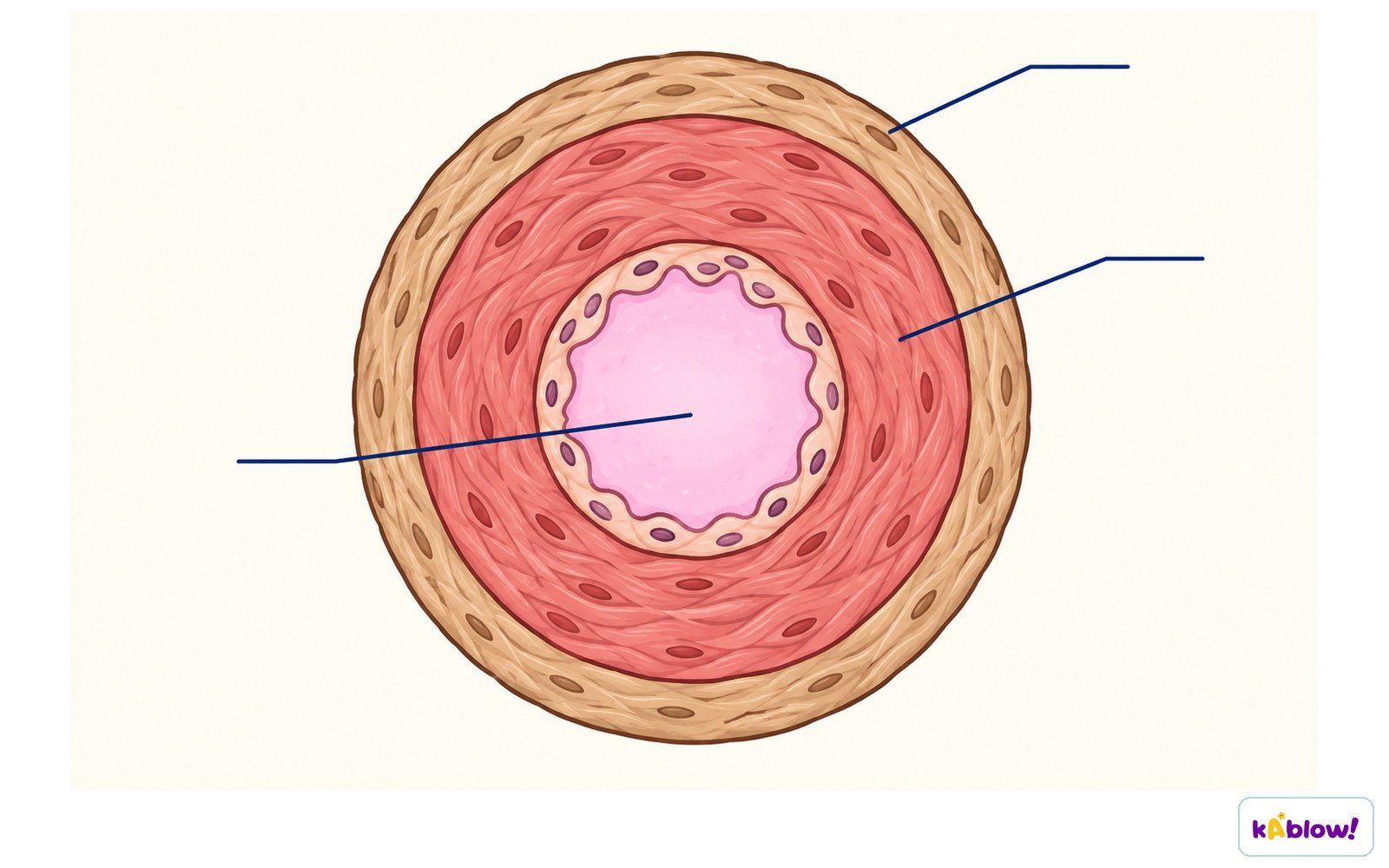
- Aan artery
- Ba capillary
- Ca vein
- Da ventricle
Unlock all 51 questions & more
Crie uma conta grátis para ver todas as perguntas, os slides, flashcards e resumos de revisão deste tópico.