Homeostasis
边玩边学
回答这些题目来赚取能量,然后钓鱼探索。无需账号。
给教育者: 面向 Homeostasis(Biology,HL)的即用型课程幻灯片, 复习笔记——用在你的课堂上,或将该知识点作为学习者可实时游玩的互动课堂活动来运行。
课程笔记
Homeostasis: Maintaining the Internal Environment
- Homeostasis is the maintenance of a constant internal environment, keeping conditions inside the body within preset limits.
- It is critically important because it maintains optimal conditions for enzyme action and cell function.
- Sensory cells detect information about conditions inside and outside the body; if conditions change, the body responds to keep them constant.
- Physiological factors controlled by homeostasis in mammals include core body temperature, blood pH, blood glucose concentration, and the osmotic concentration of the blood.
The negative feedback cycle
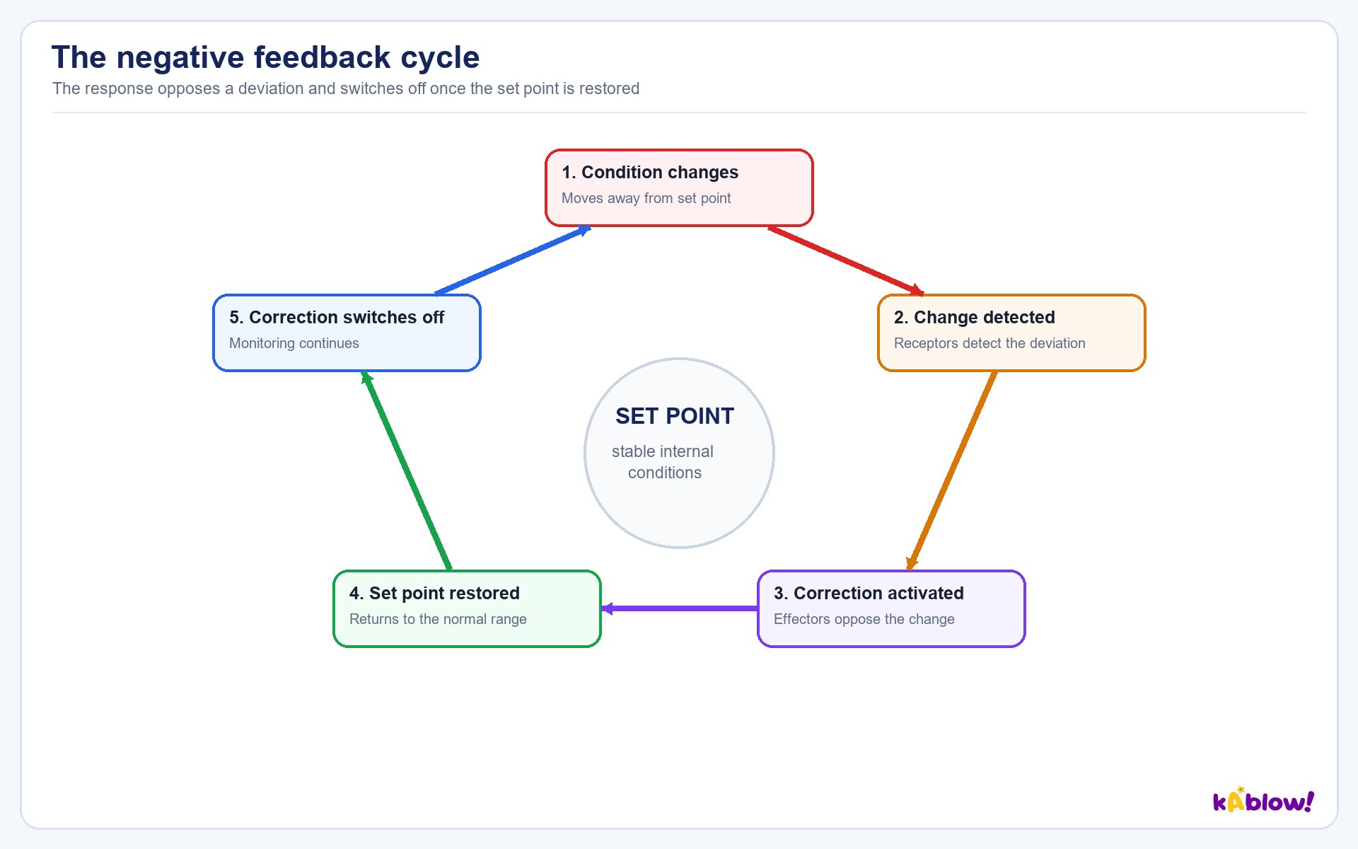
Negative Feedback Loops
- Most homeostatic control mechanisms use negative feedback loops, which work to return values to a set point by reversing the effects of any change.
- Negative feedback is essential for maintaining conditions within set limits; positive feedback instead amplifies any change.
- A negative feedback control loop involves a receptor (detects change), a coordination system (brain and nervous system transfer information), and an effector (muscles and glands bring about a response).
- The factor or stimulus is continuously monitored: if it increases, the body responds to make it decrease; if it decreases, the body responds to make it increase.
Regulation of Blood Glucose
- Blood glucose concentration must be kept within narrow limits because glucose is essential for respiration and, being soluble, affects the osmotic balance between cells and blood.
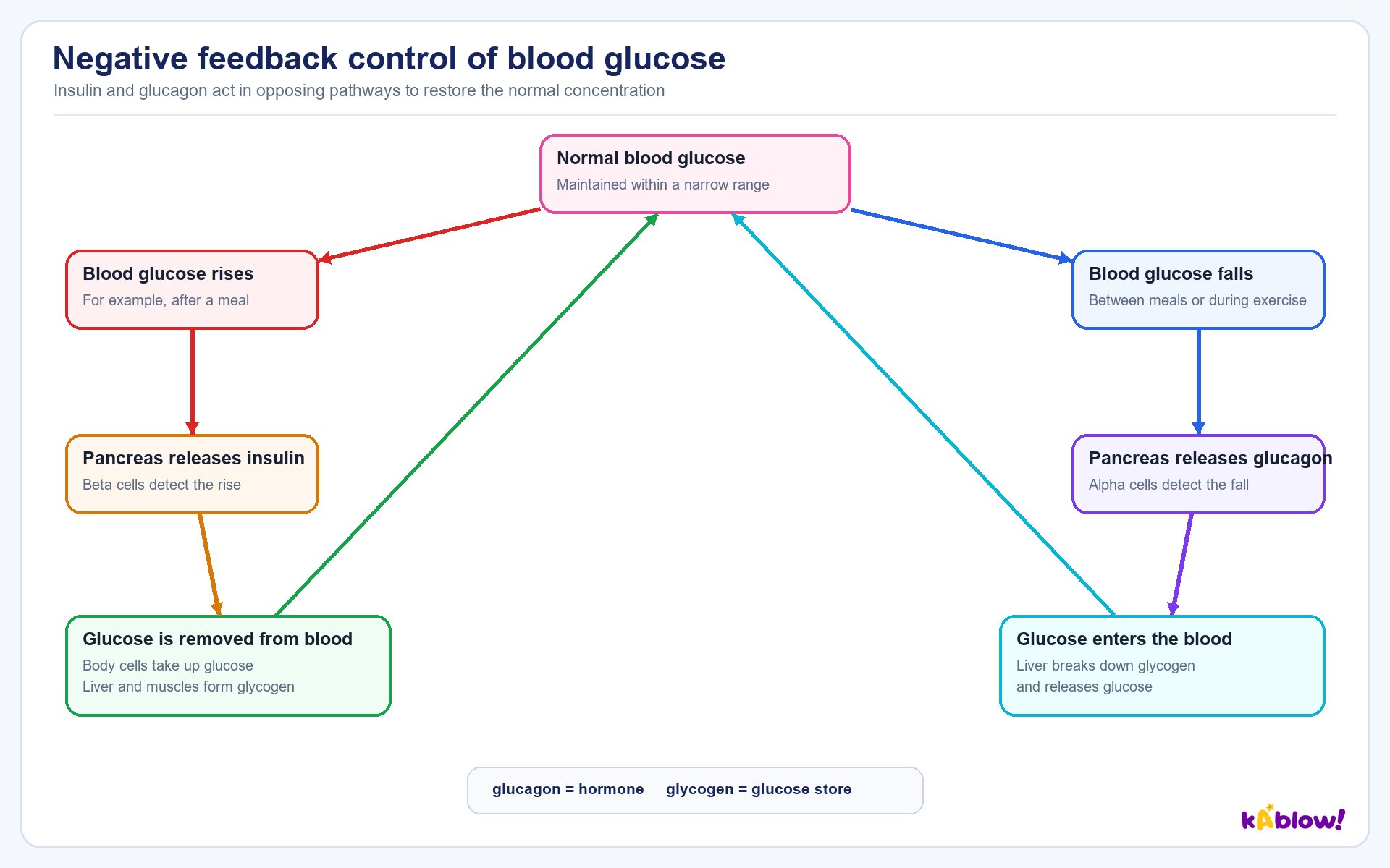
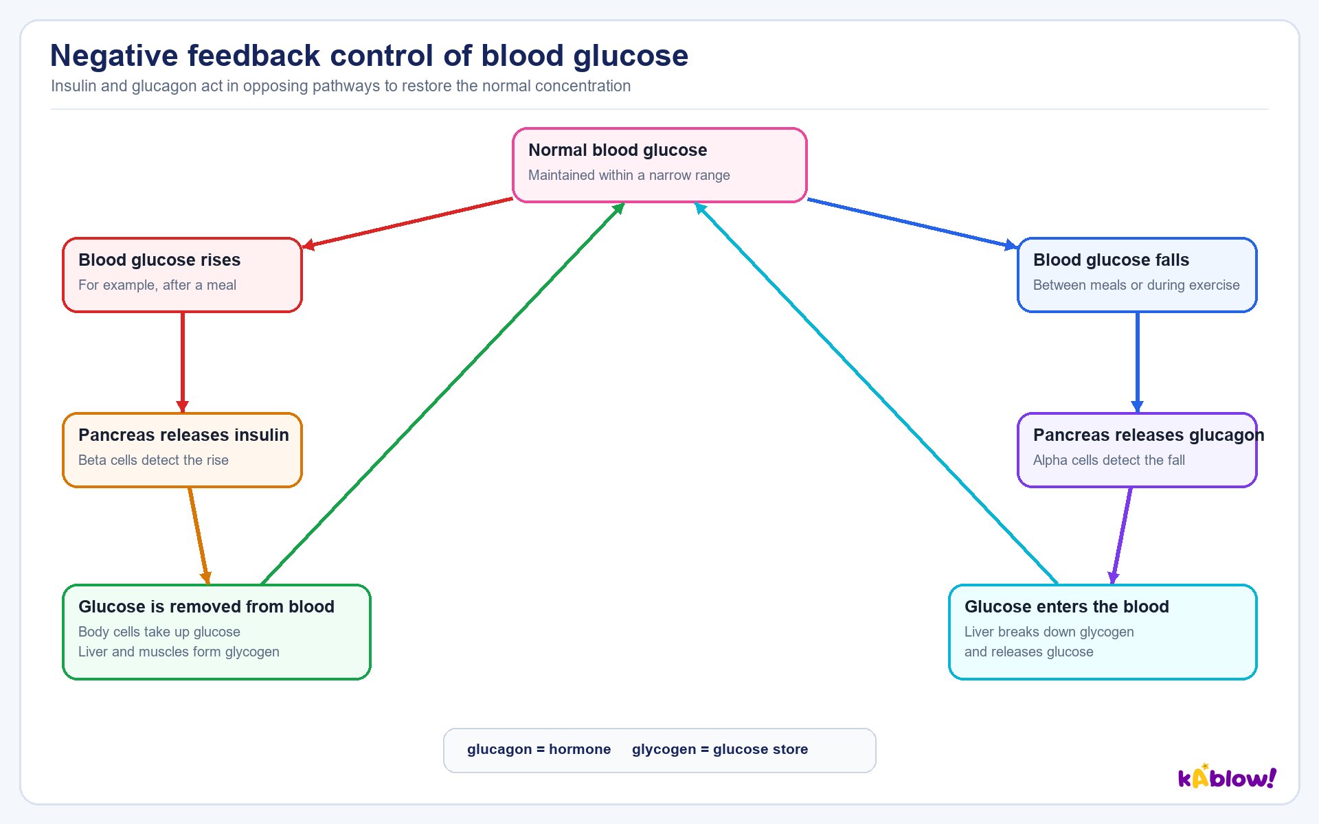
- Blood glucose is controlled by two hormones secreted by the islets of Langerhans in the pancreas: α cells secrete glucagon and β cells secrete insulin.
- The islets of Langerhans form the endocrine tissue of the pancreas; the exocrine tissue produces digestive enzymes.
- Insulin is secreted when blood glucose rises; it lowers blood glucose by opening glucose channels for facilitated diffusion into cells, converting excess glucose to glycogen (glycogenesis), increasing the rate of respiration, and converting glucose to fatty acids for storage.
- Glucagon is secreted when blood glucose falls (e.g., after fasting or exercise); it raises blood glucose by activating enzymes that hydrolyse glycogen to glucose (glycogenolysis), decreasing the rate of respiration, and converting amino acids to glucose (gluconeogenesis).
- Insulin's main target cells are in the liver and muscles; glucagon targets liver and muscle cells.
- Remember: glucagon is the hormone, while glycogen is the storage polysaccharide of animal cells.
Control of blood glucose
Diabetes: Type 1 and Type 2
- Diabetes is a condition in which the homeostatic control of blood glucose has failed or deteriorated, allowing blood glucose concentration to rise.
- The kidneys cannot filter out excess glucose, so it often appears in the urine; the increased glucose concentration causes the kidneys to produce large volumes of urine, making the individual feel thirsty due to dehydration.
- Glucose remains in the blood rather than entering cells, so cellular respiration is reduced, resulting in fatigue; dangerously high blood glucose after a meal can cause organ damage.
- Type 1 diabetes occurs when the pancreas fails to produce sufficient insulin; it normally begins in childhood due to an autoimmune response attacking the β cells of the islets of Langerhans.
- Type 1 diabetes is treated with regular blood tests, insulin injections, and a modified diet (e.g., reduced carbohydrate intake).
- Type 2 diabetes is more common and usually develops in older adults; the pancreas still produces insulin but cell membrane receptors have reduced in number or no longer respond, described as insulin resistance.
- Type 2 diabetes is managed with medication to lower blood glucose, a low-carbohydrate diet, and an exercise regime that lowers blood glucose; obesity is a major risk factor.
Opposing actions of insulin and glucagon
Thermoregulation
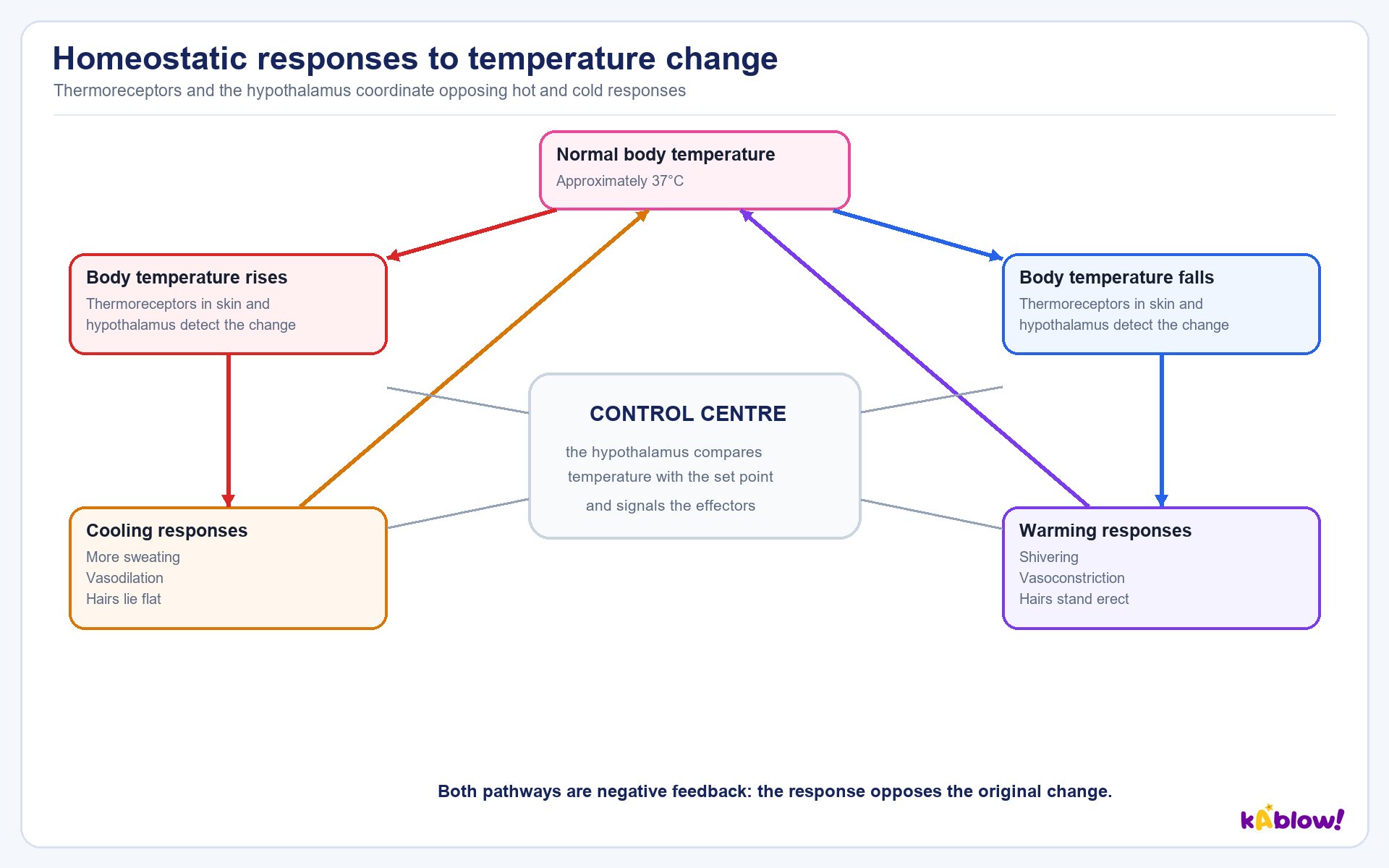
- Thermoregulation is the control of internal body temperature and is an example of negative feedback; responses reverse any deviation from pre-set limits.
- Peripheral thermoreceptors in the skin monitor external body temperature, while receptors inside the hypothalamus monitor internal body temperature.
- A stable core temperature of about 37 °C is vital for enzyme activity; lower temperatures slow reactions, while too high temperatures cause enzymes to denature.
- Endotherms such as mammals and birds maintain a constant internal body temperature using physiological mechanisms (e.g., shivering, altered metabolism) and behavioural mechanisms (e.g., seeking shade, sunbathing).
- Responses to an increase in temperature include vasodilation (arteriole muscles relax, more blood flows through skin capillaries, more heat lost by radiation), sweating (cools skin by evaporation), and flattening of hairs (allows air to circulate over skin).
- Responses to a decrease in temperature include vasoconstriction (arteriole muscles contract, less blood flows through skin capillaries, blood diverted through shunt vessels), erection of hairs (traps insulating air), shivering (rapid muscle contraction releases heat), and uncoupled respiration in brown adipose tissue (releases heat without ATP production).
- The hormone thyroxine from the thyroid gland increases the basal metabolic rate (BMR), increasing heat production.
- Vasodilation and vasoconstriction are caused by relaxing and contracting muscles in the arterioles, not the capillaries; capillaries do not have muscles in their walls.
Responses to a change in body temperature
Osmoregulation & Excretion
- The kidney has two roles: excretion and osmoregulation.
- Excretion is the process by which toxic waste products of metabolism are removed from the body; the kidneys excrete nitrogenous waste from the breakdown of excess dietary amino acids and nucleic acids.
- Nitrogenous waste is first converted into ammonia, which is highly toxic and must be removed quickly; some organisms convert ammonia into less toxic urea, which is diluted with water to form urine.
- Osmoregulation is the maintenance of a safe balance of water and solutes in cells; failure means cells could take on water and burst, or lose water and shrink due to osmosis.
- Cells with a lower water potential than their surroundings gain water by osmosis and may burst (plant cells are protected by their cell walls); cells with a higher water potential lose water and shrink.
- The units for osmotic concentration are osmoles per litre (osmol L⁻¹).
The structure of a human kidney
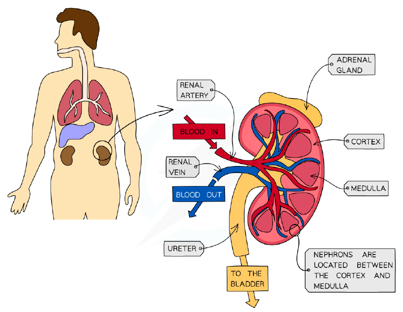
Production of Urine: Kidney Structure
- Humans have two kidneys, which remove waste products and maintain the blood's balance of water and solutes.
- The renal artery supplies oxygenated blood (containing urea and salts) to the kidneys; the renal vein carries deoxygenated blood (with urea and excess salts removed) away.
- The ureter carries urine from each kidney to the bladder, which stores urine temporarily; the urethra releases urine outside the body.
- The kidney is surrounded by a fibrous capsule and has three main regions: the cortex, the medulla, and the renal pelvis.
- Each kidney contains thousands of nephrons, the functional units responsible for urine formation.
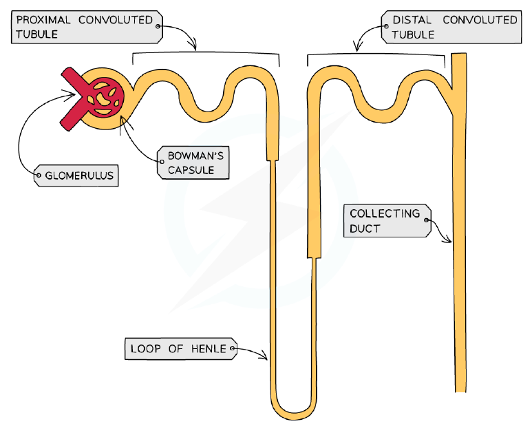
- The cortex contains the glomerulus, Bowman's capsule, proximal convoluted tubule, and distal convoluted tubule; the medulla contains the loop of Henle and collecting duct; all nephrons drain into the renal pelvis, which connects to the ureter.
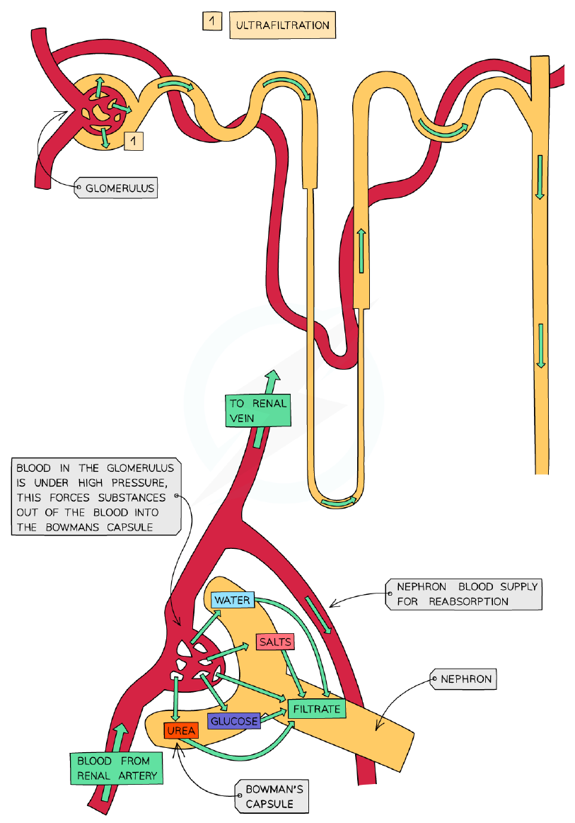
Ultrafiltration and Selective Reabsorption
- Each glomerulus is supplied by an afferent arteriole and drained by an efferent arteriole; the afferent arteriole is wider than the efferent arteriole, increasing blood pressure in the glomerulus.
- Ultrafiltration occurs as high blood pressure forces small molecules from the glomerulus into the Bowman's capsule, forming glomerular filtrate; these molecules include chloride ions, sodium ions, glucose, urea, and amino acids.
- Large molecules such as proteins remain in the blood and do not pass into the filtrate; red and white blood cells and platelets also remain in the blood.
- The glomerular filtrate passes through three layers: the capillary endothelium (with fenestrations), the basement membrane (a mesh of collagen and glycoproteins acting as a sieve), and the Bowman's capsule epithelium (podocytes with gaps between projections).
- Selective reabsorption reabsorbs useful substances (water, salts, glucose, amino acids) into the blood as the filtrate passes along the nephron; most occurs in the proximal convoluted tubule.
- The proximal convoluted tubule epithelium is adapted by microvilli (increase surface area), co-transporter proteins (transport specific solutes), many mitochondria (provide energy for Na⁺-K⁺ pumps), and tightly packed cells (no fluid passes between cells).
- In selective reabsorption, sodium ions are actively transported out, chloride ions follow by diffusion, sugars and amino acids are transported by co-transporter proteins, water leaves by osmosis, and urea moves out by diffusion.
Ultrafiltration
Water Reabsorption in the Loop of Henlé and Collecting Duct
- The loop of Henlé enables production of urine more concentrated than blood, conserving water; it can also produce urine less concentrated than blood when water intake is high.
- In the ascending limb, sodium and chloride ions are pumped out into the medulla, lowering its water potential; the ascending limb is impermeable to water.
- The descending limb is permeable to water but has few transport proteins, so water moves out by osmosis and the filtrate's water potential decreases as it descends.
- The low water potential in the medulla created by the ascending limb also enables water reabsorption from the collecting duct by osmosis.
- The vasa recta is the capillary flowing alongside the loop of Henlé; it also supplies oxygen and removes carbon dioxide from the respiring cells.
- Osmoregulation is an example of homeostasis; the kidneys alter the amount of water reabsorbed by changing the permeability of the distal convoluted tubule and collecting duct.
- ADH (antidiuretic hormone) is released from the posterior pituitary gland, regulated by the hypothalamus, which monitors blood composition via osmoreceptor cells.
Structure of a nephron
ADH and Changes in Blood Supply to Organs
- Low blood water content (high blood solute concentration) is detected by the hypothalamus, which causes the pituitary to secrete ADH; ADH increases the permeability of the distal convoluted tubule and collecting duct to water.
- ADH increases permeability by increasing the number of aquaporin channel proteins in the cell surface membranes; vesicles containing aquaporins fuse with the membrane.
- More water is reabsorbed into the blood, so blood water content increases and a small volume of concentrated urine is produced.
- High blood water content (low blood solute concentration) leads to less ADH release; fewer aquaporins are present, less water is reabsorbed, and a large quantity of dilute urine is produced.
- The circulatory system diverts blood flow to different organs by vasodilation or vasoconstriction in the arterioles supplying capillary beds.
- Blood flow to skeletal muscles is low during sleep, increases during wakefulness, and increases greatly during exercise; blood flow to the gut increases after a meal and decreases during exercise.
- Blood flow to the brain remains relatively constant regardless of activity level (increasing slightly during REM sleep); blood flow to the kidneys does not change significantly with activity, increasing slightly during sleep and rest and decreasing slightly during prolonged exercise.
幻灯片
练习题
免费预览——51 题中的 8 题。注册即可查看全部。
1.What is the term for the process of maintaining a constant internal environment within preset limits?
Easy- AHomeostasis
- BOsmoregulation
- CThermoregulation
- DExcretion
2.Which of the following is NOT a physiological factor controlled by homeostasis in mammals?
Easy- ACore body temperature
- BBlood pH
- CBlood glucose concentration
- DBlood group
3.Negative feedback mechanisms work to return values to a set point by reversing the effects of any change.
EasyTrue or false?
4.Which component of a negative feedback loop detects a change in a physiological factor?
Easy- AEffector
- BReceptor
- CCoordination system
- DSet point
5.Which of the following are examples of physiological factors controlled by homeostasis in mammals? (select all that apply)
Medium- ACore body temperature
- BBlood pH
- CBlood glucose concentration
- DBlood group
- EOsmotic concentration of the blood
6.Match each component of a negative feedback loop with its role.
Medium- Receptor
- Coordination system
- Effector
- Detects change in a physiological factor
- Transfers information between parts of the body
- Brings about a response
7.Which cells in the islets of Langerhans secrete insulin?
Easy- Aα cells
- Bβ cells
- CAcinar cells
- DPodocytes
8.What is the effect of insulin on blood glucose concentration?
Medium- AIt increases blood glucose concentration
- BIt decreases blood glucose concentration
- CIt has no effect on blood glucose concentration
- DIt only affects blood glucose during exercise
历年真题
该知识点的历年真题练习即将推出。
即将推出