Gas Exchange
邊玩邊學
回答這些題目賺取能量,接著就能釣魚、探索。不需要帳號。
給教育者: 為 Gas Exchange(Biology、HL)準備好可直接使用的課程投影片, 複習筆記——用於你的課程,或把這個主題當成互動班級活動,讓學習者以即時遊戲的方式進行。
課程筆記
Gas Exchange in Organisms
- Cellular respiration releases energy as ATP by oxidising substrates such as glucose; aerobic respiration requires oxygen and produces carbon dioxide as a waste product.
- Gas exchange is the diffusion of oxygen into an organism and carbon dioxide out of it, between the organism and its environment.
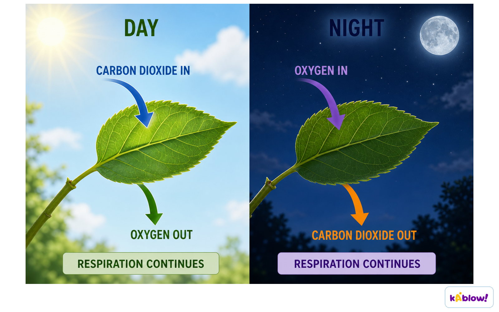
- In plants, carbon dioxide is absorbed and oxygen is released during the day as a result of photosynthesis.
- The rate of diffusion depends on: surface area (larger surface = faster diffusion), concentration gradient, and diffusion distance (shorter distance = faster diffusion).
- Small unicellular organisms such as amoeba have a large surface area to volume ratio and a short diffusion distance, so diffusion alone is sufficient for gas exchange.
- As organisms increase in size, the surface area to volume ratio decreases and the diffusion distance increases, so diffusion alone cannot supply every cell with oxygen.
- Large, active organisms have higher metabolic demands and require specialised gas exchange organs; their external surface is adapted for protection, not gas exchange.
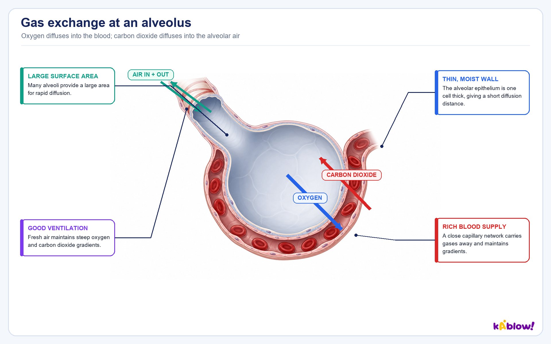
An alveolus and its capillary
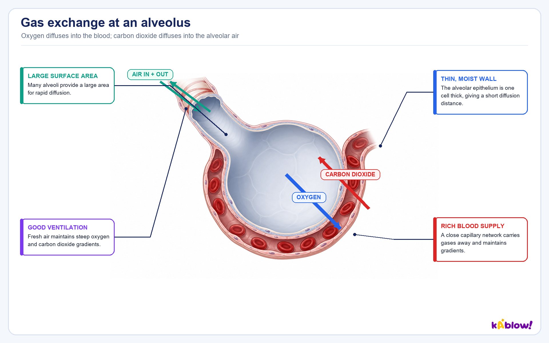
Properties of Gas Exchange Surfaces
- Gas exchange surfaces must be permeable so gases can move across them.
- They must be thin to provide a short diffusion distance for oxygen and carbon dioxide.
- They must be moist so that gases can dissolve before diffusing.
- They must have a large surface area so many gas molecules can diffuse at the same time.
- A steep concentration gradient is maintained by a dense network of blood vessels, continuous blood flow, and ventilation with air (lungs) or water (gills).
- Blood acts as a good transport medium, constantly carrying oxygen away from and carbon dioxide towards the gas exchange surface.
Features of gas exchange surfaces
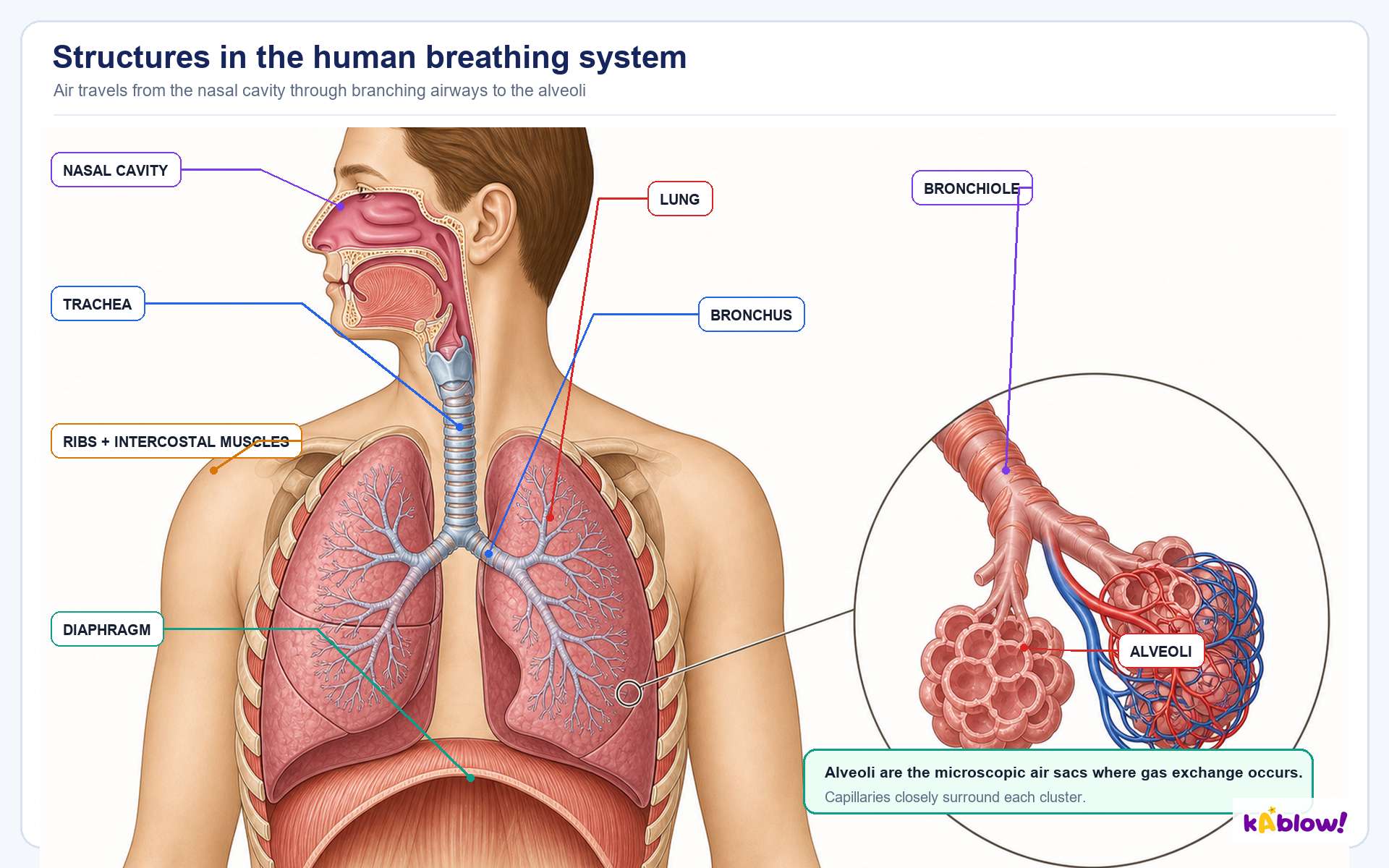
Mammalian Lungs: Adaptations
- Air enters through the nose and mouth, then travels down the trachea, which is supported by rings of cartilage to keep it open while allowing movement and flexibility.
- The trachea divides into two bronchi; their walls contain cartilage and smooth muscle that can contract or relax to change airway diameter.
- The trachea and bronchi are lined with ciliated epithelium to remove particles trapped in mucus.
- Bronchioles branch off the bronchi and have smooth muscle in their walls to regulate airflow by dilating or constricting.
- Alveoli are grouped at the ends of bronchioles, providing a large surface area for gas exchange, and are surrounded by an extensive capillary network.
- Deoxygenated blood enters the capillary beds from the pulmonary artery and oxygenated blood leaves via the pulmonary vein, maintaining the concentration gradient.
- The alveolar wall is a single layer of epithelial cells; Type I pneumocytes are thin for rapid diffusion, while Type II pneumocytes secrete surfactant that lowers surface tension and prevents alveoli from collapsing and sticking together during expiration.
The human breathing system
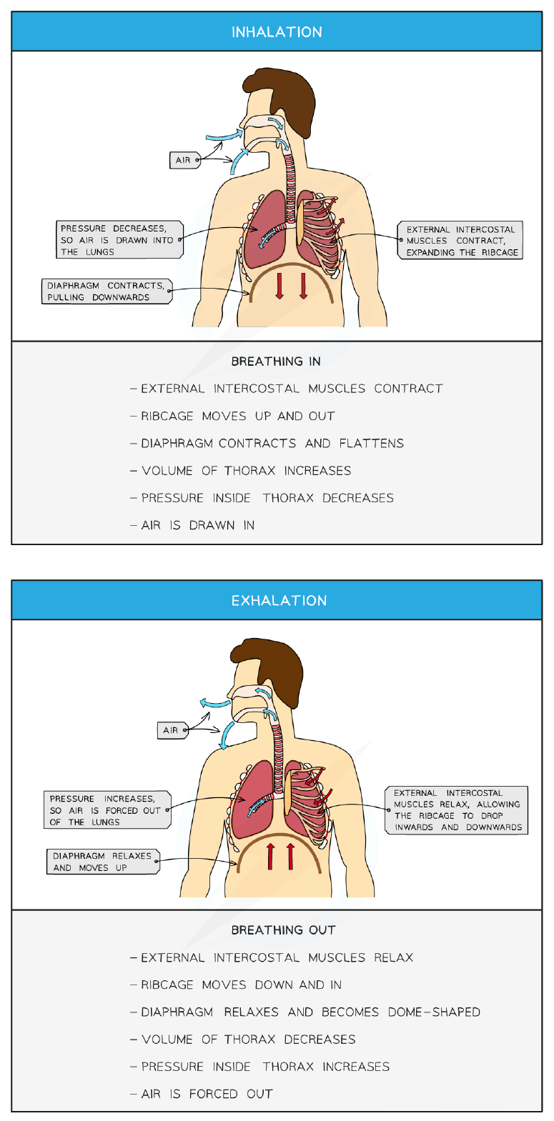
Mechanism of Ventilation
- Ventilation replaces older air in the lungs with fresh air, maintaining the concentration gradient of oxygen and carbon dioxide between the alveoli and blood.
- Inspiration (breathing in) increases chest volume and decreases air pressure below atmospheric pressure, so air rushes in down the pressure gradient.
- During inspiration, the diaphragm contracts and flattens and the external intercostal muscles contract, moving the ribcage upwards and outwards.
- Expiration (breathing out) is mainly passive: the external intercostal muscles relax, the diaphragm relaxes and becomes dome-shaped, and elastic fibres in the alveoli walls recoil, reducing lung volume.
- Active expiration occurs when excess air must be expelled: internal intercostal muscles contract to pull the ribs down and in, and abdominal muscles contract to push organs upwards against the diaphragm.
- The diaphragm and external intercostal muscles are an antagonistic pair; the internal intercostal muscles and external intercostal muscles are another antagonistic pair involved in ventilation.
Inhalation and exhalation
Measuring Lung Volumes: Skills
- A spirometer contains a chamber filled with water covered by a hinged plastic lid; the person breathes through a mouthpiece connected to the chamber, and the lid moves up and down as breathing occurs.
- The spirometer chamber can be filled with air to determine lung capacity, or with oxygen and soda lime (to absorb carbon dioxide) to measure oxygen consumption.
- Spirometer traces are created by drawing a line on a revolving drum or by a computer that draws a graph of the results.
- Tidal volume is the volume of air inhaled and exhaled during normal breathing; exercise increases tidal volume.
- Inspiratory reserve volume is the difference between the maximum inspiratory level and tidal volume; expiratory reserve volume is the difference between the maximum expiratory level and tidal volume.
- Vital capacity is the total amount of air exhaled after a deep breath, calculated as VC = TV + IRV + ERV.
- Ventilation rate is the number of inhalations or exhalations per minute; exercise increases the ventilation rate.
Gas Exchange in Plants
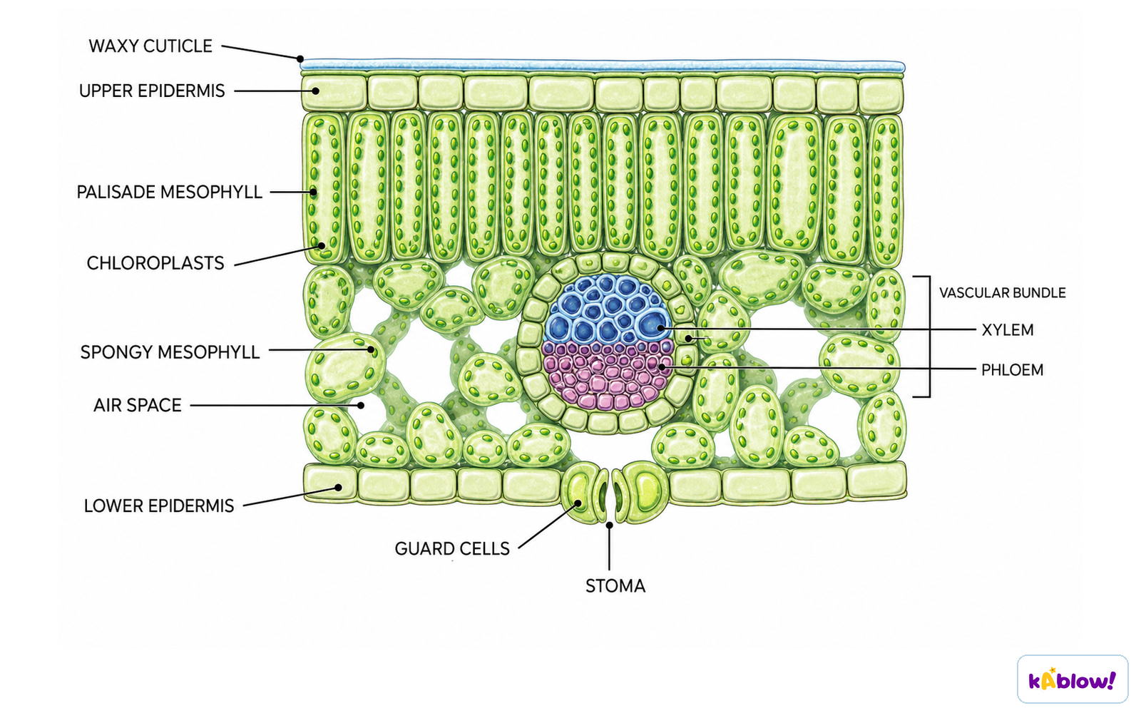
- The leaf contains epidermal tissue (outer boundary), mesophyll tissue (bulk of internal structure), and vascular tissue (transport).
- The lower epidermis contains stomata (singular stoma), each surrounded by two guard cells that control opening and closing.
- Guard cells become turgid when water moves in, changing shape to open the stomata; they become flaccid when water is lost, causing stomata to close.
- Stomata allow diffusion of oxygen and carbon dioxide into and out of the leaf; the epidermis is covered by a waxy cuticle that forms an impermeable barrier.
- Palisade mesophyll lies beneath the upper epidermis and contains many chloroplasts for maximum photosynthesis; spongy mesophyll contains large air spaces for gas exchange.
- Xylem transports water and mineral ions from roots to leaves; phloem transports the products of photosynthesis from leaves to other parts of the plant.
- Leaf adaptations for gas exchange include: waxy cuticle (controls gas exchange and water loss), stomata mostly in lower epidermis (reduces water loss), air spaces (maintain concentration gradient), spongy mesophyll (increases surface area), guard cells (control gas exchange and water loss), and veins (bring water for photosynthesis and transpiration).
Gas exchange in plants, day and night
Transpiration: Consequence of Gas Exchange
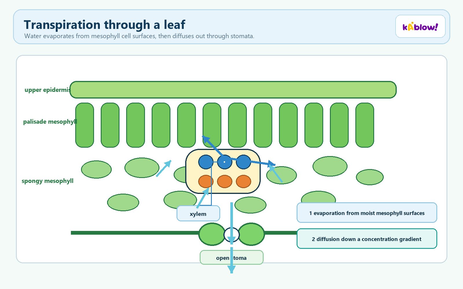
- Transpiration is the loss of water vapour from leaves through stomata; it is an inevitable consequence of gas exchange because stomata must be open for photosynthesis.
- Transpiration provides cooling via evaporation, helps uptake of mineral ions via the transpiration stream, and provides turgor pressure for support of leaves and non-woody stems.
- Air movement: more air movement increases transpiration by carrying water vapour away, increasing the concentration gradient.
- Temperature: higher temperatures increase transpiration up to a point, as increased kinetic energy speeds evaporation; if too high, stomata close to prevent excess water loss.
- Light intensity: higher light intensity increases transpiration up to a point because stomata open in light for gas exchange; once all stomata are open, further increase has no effect.
- Humidity: higher humidity reduces transpiration because the air is saturated with water vapour, reducing the concentration gradient; at equilibrium there is no net loss of water vapour.
- A potometer measures the rate of water uptake, which is used to represent the rate of transpiration; bubble potometers measure air bubble movement, mass potometers measure change in mass.
Transpiration through a leaf
Drawing Leaf Structure: Skills
- You must be able to identify: chloroplasts, cuticle, guard cells, stomata, upper and lower epidermis, palisade mesophyll, spongy mesophyll, air spaces, and vascular bundles (xylem and phloem).
- Plan diagrams are drawings made from micrographs or specimens under low magnification; no individual cells are drawn, only tissue layers enclosed by lines.
- Use a sharp pencil and draw clear, continuous lines; do not shade any part of the drawing.
- Make sure proportions and observations are accurate; draw what you actually see, not what you expect from a textbook.
- Draw large enough to fill at least half the available space.
- When labelling: use a ruler for label lines, avoid arrowheads, ensure lines stop at the structure, do not cross label lines, and write all labels horizontally.
Leaf cross-section
Determining Stomatal Density: Skills
- Stomatal density is the number of stomata per unit area; it can indicate a plant's likely response to dry weather or predict behaviour in windy or wet climates.
- Method: paint clear nail varnish on the underside of a leaf, let it dry, peel off with sellotape, place the leaf cast on a microscope slide, and count stomata in the field of view.
- Count at least 3 separate fields of view and take a mean value to eliminate anomalous results and calculate a reliable mean.
- Use a stage micrometer at the same magnification to measure the diameter of the field of view, then calculate the radius and area (Area = πr²).
- Calculate density by dividing the mean number of stomata by the area of the field of view; give units in stomata per mm².
- Limitations: not all species create strong imprints, solvent-based nail varnish can destroy cell structure, and water-based varnish dries more slowly.
- Repeating measurements increases the reliability of quantitative data; anomalous measurements (outliers) are omitted when calculating the mean.
Haemoglobin & Oxygen
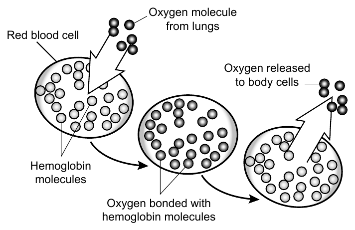
- Haemoglobin is a globular protein found in red blood cells; it consists of four polypeptide subunits, each with an iron-containing haem group that can bind one oxygen molecule, so each haemoglobin transports four oxygen molecules.
- The pressure exerted by each gas in a mixture is its partial pressure (p); the partial pressure of oxygen is denoted pO₂.
- The first oxygen molecule binds slowly due to the shape of haemoglobin; after it binds, the protein changes shape (conformation) making it easier for the next oxygen molecules to bind — this is cooperative binding.
- Affinity describes how easily haemoglobin binds and dissociates with oxygen; at high pO₂ (alveoli) affinity is high, and at low pO₂ (respiring muscle) affinity is low.
- Foetal haemoglobin has a higher affinity for oxygen than adult haemoglobin, allowing the foetus to obtain oxygen from the mother's blood at the placenta where pO₂ is low.
- On the oxygen dissociation curve, foetal haemoglobin shifts to the left of adult haemoglobin, meaning it has a higher percentage saturation at any given pO₂.
- Carbon dioxide is an allosteric inhibitor of haemoglobin; when it binds, it is more difficult for oxygen to bind, lowering haemoglobin's affinity for oxygen.
- Carbon dioxide has less of an allosteric effect on foetal haemoglobin, enabling it to maintain a higher affinity for oxygen even when carbon dioxide is bound.
Haemoglobin carries oxygen
The Bohr Shift
- The Bohr effect (Bohr shift) describes changes in the oxygen dissociation curve caused by carbon dioxide levels.
- When the partial pressure of carbon dioxide in the blood is high, haemoglobin's affinity for oxygen is reduced; this occurs in respiring tissues producing carbon dioxide.
- Carbon dioxide lowers the pH of the blood: CO₂ combines with water to form carbonic acid, which dissociates into hydrogen carbonate ions and hydrogen ions.
- Hydrogen ions bind to haemoglobin, causing the release of oxygen.
- This is helpful because haemoglobin gives up oxygen more readily in respiring tissues where it is needed.
- On a graph, the dissociation curve shifts to the right when CO₂ levels increase, meaning at any given pO₂ the percentage saturation of haemoglobin is lower at higher CO₂ levels.
The Oxygen Dissociation Curve
- The oxygen dissociation curve shows the rate at which oxygen associates with and dissociates from haemoglobin at different partial pressures of oxygen (pO₂).
- Haemoglobin is saturated when all four oxygen binding sites are occupied.
- At low pO₂ (bottom left), oxygen binds slowly; haemoglobin has a low affinity and saturation is low.
- At medium pO₂ (central region), oxygen binds more easily and saturation increases quickly; a small increase in pO₂ causes a large increase in saturation.
- At high pO₂ (top right), oxygen binds easily and haemoglobin becomes saturated; a large increase in pO₂ has only a small effect on saturation because most binding sites are occupied.
- Reading from right to left: in the lungs (high pO₂) there is very little dissociation; at medium pO₂ oxygen dissociates readily (steep region), corresponding to respiring tissues; at low pO₂ dissociation slows again.
- The curve's shape is explained by: slow binding of the first oxygen molecule (shallow bottom left), cooperative binding speeding up subsequent binding (steeper middle), and difficulty binding the fourth oxygen molecule as saturation approaches (levelling off top right).
投影片
練習題
免費預覽——61 題中的 8 題。註冊即可查看全部。
1.Gas exchange in living organisms takes place by which process?
Easy- ADiffusion
- BOsmosis
- CActive transport
- DMass flow
2.Which adaptation of a gas exchange surface reduces the diffusion distance?
Easy- AA thin tissue layer
- BA large surface area
- CA moist surface
- DA dense network of blood vessels
3.What is the main role of cartilage in the trachea and bronchi?
Easy- ATo support the airway and keep it open
- BTo trap dust particles
- CTo produce mucus
- DTo control the diameter of the airway
4.Which of the following is a function of the fluid secreted by Type II pneumocytes?
Medium- AIt reduces surface tension to prevent alveolar collapse
- BIt increases surface tension to keep alveoli open
- CIt traps dust particles in the alveoli
- DIt transports oxygen across the alveolar wall
5.What is the correct term for the volume of air inhaled and exhaled during normal breathing?
Medium- ATidal volume
- BVital capacity
- CInspiratory reserve volume
- DExpiratory reserve volume
6.Which structure in a leaf controls the opening and closing of stomata?
Easy- AGuard cells
- BEpidermal cells
- CMesophyll cells
- DXylem vessels
7.How does an increase in carbon dioxide concentration affect the oxygen dissociation curve of adult haemoglobin?
Medium- AIt shifts the curve to the right, reducing haemoglobin's affinity for oxygen
- BIt shifts the curve to the left, increasing haemoglobin's affinity for oxygen
- CIt has no effect on the curve
- DIt makes the curve linear
8.In the oxygen dissociation curve, what does a shift to the left indicate?
Medium- AA higher affinity of haemoglobin for oxygen
- BA lower affinity of haemoglobin for oxygen
- CA higher partial pressure of carbon dioxide
- DA lower percentage saturation of haemoglobin at any given pO2
歷屆試題
這個主題的歷屆試題練習即將推出。
即將推出